
News & Announcements
Biomarker Standards for Fabry Disease Diagnosis and Monitoring
Article from 2019-08-01
This article was originally published in the August 2019 edition of Matreya’s Newsletter for Glyco/Sphingolipid Research (PDF).
Lysosomal storage disorders are a set of more than 70 inherited conditions that result in the accumulation of various lipids in cells due to an inability to enzymatically degrade them. Fabry disease (FD) is one such disorder that is characterized by a deficiency of the enzyme α-galactosidase A, resulting in an accumulation of globotriaosylceramide (Gb3), globotriaosylsphingosine (lyso-Gb3), galabiaosylceramide (Ga2), and blood group B glycolipids. This debilitating disease is an X-linked chromosomal disorder and early diagnosis is critical as progression will lead to multiorgan dysfunction and early death. To aid in timely detection and treatment monitoring, several disease biomarkers have been identified, including Gb3 and lyso-Gb3.
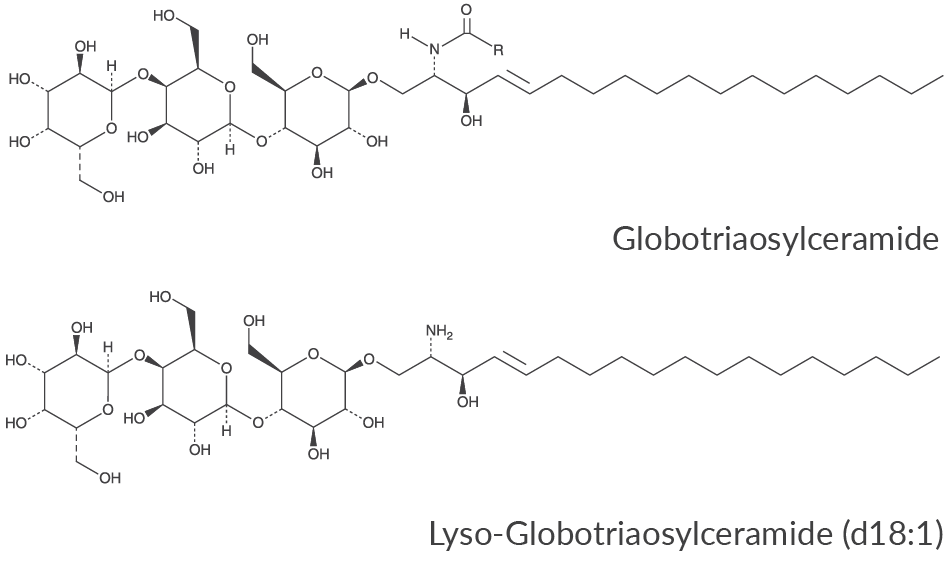
The use of lyso-Gb3 for the determination of Fabry disease has now been well established, resulting in the development of reproducible and highly sensitive methods that require extremely small plasma, dried blood spot, or urine samples.1,2 Lyso-Gb3 has been demonstrated to be an effective biomarker for FD in symptomatic patients, showing higher diagnostic sensitivity than Gb3.1 However, this lipid has not always been found to be a good candidate biomarker for asymptomatic females.3 Heterozygous females can also manifest symptoms of FD making early detection of critical importance in these cases as well. Urinary Gb3 (but not plasma Gb3) has been reported to be elevated in both symptomatic and asymptomatic males and females even though its sensitivity is lower than lyso-Gb3. Although levels of Gb3 do not necessarily correlate with disease severity, and though false positives do occur using this biomarker, Gb3 has been recognized as a useful diagnostic marker and may also indicate the formation of antibodies during enzyme replacement therapy (ERT).3 Other possible biomarkers for FD include galabiaosylceramide (Ga2)4 and blood group B glycolipids.
Currently FD is most often treated by ERT and the levels of Gb3 and lyso-Gb3 have also been found to be useful in monitoring the levels of replacement enzymes needed as well as disease progression. ERT has shown a reduction not only in plasma Gb3 but also in plasma lyso-Gb3. Because FD is a result of deficient enzyme activity, ERT is able to successfully halt the disease progress in many patients. This is especially evident in patients that begin treatment early in the disease progression as untreated patients can quickly develop irreversible organ damage.5
Internal standards for Gb3 and lyso-Gb3 are critical for the effective quantitation of these FD biomarkers. Glycinated lyso-Gb3 was explored as an internal standard, and the physical and chemical properties were found to be almost identical to that of natural lyso-Gb3 in terms of extraction, stability, and sensitivity, making it an excellent internal standard for clinical work.6 This internal standard contains a glycine molecule attached to the amine of lyso-Gb3, preserving the key primary amine functionality. Another commonly used internal standard for lyso-Gb3 analysis is lactosylsphingosine, which lacks the terminal galactose of lyso-Gb3 but does contain a primary amine.2,6 For Gb3 analysis, the most useful internal standard is a stable isotope-labeled standard (such as deuterated octadecanoyl-Gb3) or a well-defined Gb3 containing an unusual fatty acid (such as heptadecanoyl-Gb3). Armed with these internal standards, diagnosis and monitoring of FD can go forward steadily.
An example of the ongoing development of sensitive and efficient analyses of lyso-Gb3 in FD is seen in the work by Lukas and coworkers.2 Three 3.2 mm dried blood spot samples were extracted with DMSO:water 1:1 in the presence of lyso-Gb2 (as an internal standard) in ethanol with agitation and sonication. After particle filtration by centrifugation, the sample was ready for analysis. Similarly, 25 μl of plasma was extracted in ethanol in the presence of lyso-Gb2 (again as an internal standard). After protein precipitation, the supernatant was filtrated by centrifugation and analyzed. Analysis was performed by UPLC/triple quadrupole mass spectrometer in MRM mode, monitoring the mass transitions of both lyso-Gb3 and lyso-Gb2.
Another method has been evaluated for the analysis of lyso-Gb3 from dried blood spots by Johnson and colleagues.7 This HPLC-MS/MS method yielded reproducible results in patients with Fabry disease, although the method was found to be unsuitable for newborn screening and late onset females.
A method that takes advantage of detectable lyso-Gb3 in urine is reported by Gold and coworkers and includes the use of a stable isotope-labeled lyso-Gb3 (not yet commercially available).1 Urine was extracted in methanol/chloroform along with the internal standard. The sample was centrifuged, and the supernatant diluted with chloroform/water to make an upper and lower layer. The upper methanol/water phase was collected, evaporated, and partitioned with butanol/water. The upper butanol phase was collected, evaporated, and re-dissolved in methanol for analysis by UPLC-ESI-MS/MS.
It will be interesting to see what the future holds for enhanced early diagnosis, treatment, and monitoring of FD. As methods become more refined, we hope to see techniques such as solid phase microextraction being developed for even more efficient analysis. Equipped with an expanding array of standards, researchers can continue to delve deeper into the role of accumulated lipids in this disease’s devastating pathogenesis. The recent and ongoing push to make newborn screening for α-galactosidase A more prevalent will undoubtedly have tremendous benefits for the generations ahead.
Available Globotriaosylceramides
- Globotriaosylceramides (porcine)
- Lyso-Globotriaosylceramide (d18:1)
- C16 Globotriaosylceramide (d18:1/16:0)
- C18 Globotriaosylceramide (d18:1/18:0)
- C23 Globotriaosylceramide (d18:1/23:0)
Gb3 and lyso-Gb3 Internal Standards
- C17 Globotriaosylceramide (d18:1/17:0)
- C18 Globotriaosylceramide-d3 (d18:1/18:0-d3)
- N-Glycine Globotriaosylsphingosine (d18:1)
- Lactosylsphingosine (d18:1) (synthetic)
- Lactosylsphingosine (d18:1)
References
1. Gold, H., Mirzaian, M., Dekker, N., et al. Quantification of globotriaosylsphingosine in plasma and urine of Fabry patients by stable isotope ultraperformance liquid chromatography-tandem mass spectrometry. Clin. Chem. 59(3), 547-556 (2013).
2. Lukas, J., Scalia, S., Eichler, S., et al. Functional and clinical consequences of novel α-galactosidase A mutations in Fabry disease. Hum. Mutat. 37(1), 43-51 (2016).
3. Paschke, E., Fauler, G., Winkler, H., et al. Urinary total globotriaosylceramide and isoforms to identify women with Fabry disease: A diagnostic test study. Am. J. Kidney Dis.57(5), 673-681 (2011).
4. Boutin, M. and Auray-Blais, C. Metabolomic discovery of novel urinary galabiosylceramide analogs as Fabry disease biomarkers. J. Am. Soc. Mass Spectrom. 26(3), 499-510 (2015).
5. Hopkin, R.J., Cabrera, G., Charrow, J., et al. Risk factors for severe clinical events in male and female patients with Fabry disease treated with agalsidase beta enzyme replacement therapy: Data from the Fabry Registry. Mol. Genet. Metab. 119(1-2), 151-159 (2016).
6. Krüger, R., Tholey, A., Jakoby, T., et al. Quantification of the Fabry marker lysoGb3 in human plasma by tandem mass spectrometry. J. Chromatogr. B Analyt. Technol. Biomed. Life Sci.883-884, 128-135 (2012).
7. Johnson, B., Mascher, H., Mascher, D., et al. Analysis of lyso-globotriaosylsphingosine in dried blood spots. Ann. Lab. Med.33(4), 274-278 (2013).
Contact Info
Cayman Chemical1180 East Ellsworth RoadAnn Arbor, Michigan 48108 USAToll Free: (800) 364-9897(USA and Canada only)Fax: (734) 971-3640


