
News & Announcements
Screening Mitochondrial Toxicity
Screening Mitochondrial Toxicity
Article from 2014-03-01
David L. Hoffman, Ph.D.
ETC Activity Assay KitsCreate a mitochondrial function/toxicity profile with Cayman’s simple, cost effective assay kits developed to screen potential inhibitors of the electron transport chain.
The mitochondrion, known primarily as “the powerhouse of the cell” is critical for the maintenance of cellular homeostasis. Under aerobic conditions, the majority of ATP utilized by the cell is generated by mitochondria through oxidative phosphorylation. Through the reduction of oxygen and the oxidation of nutrients obtained through metabolic reactions, the Electron Transport Chain (ETC) pumps protons from the mitochondrial matrix to the intermembrane space, establishing a proton gradient (Figure 1).1 This proton gradient is comprised of both electrochemical (ΔΨm) and pH (ΔpH) components and acts as the driving force for oxidative phosphorylation. The ATP synthase (Complex V) harnesses the energy stored in this gradient to produce ATP from ADP and inorganic phosphate.
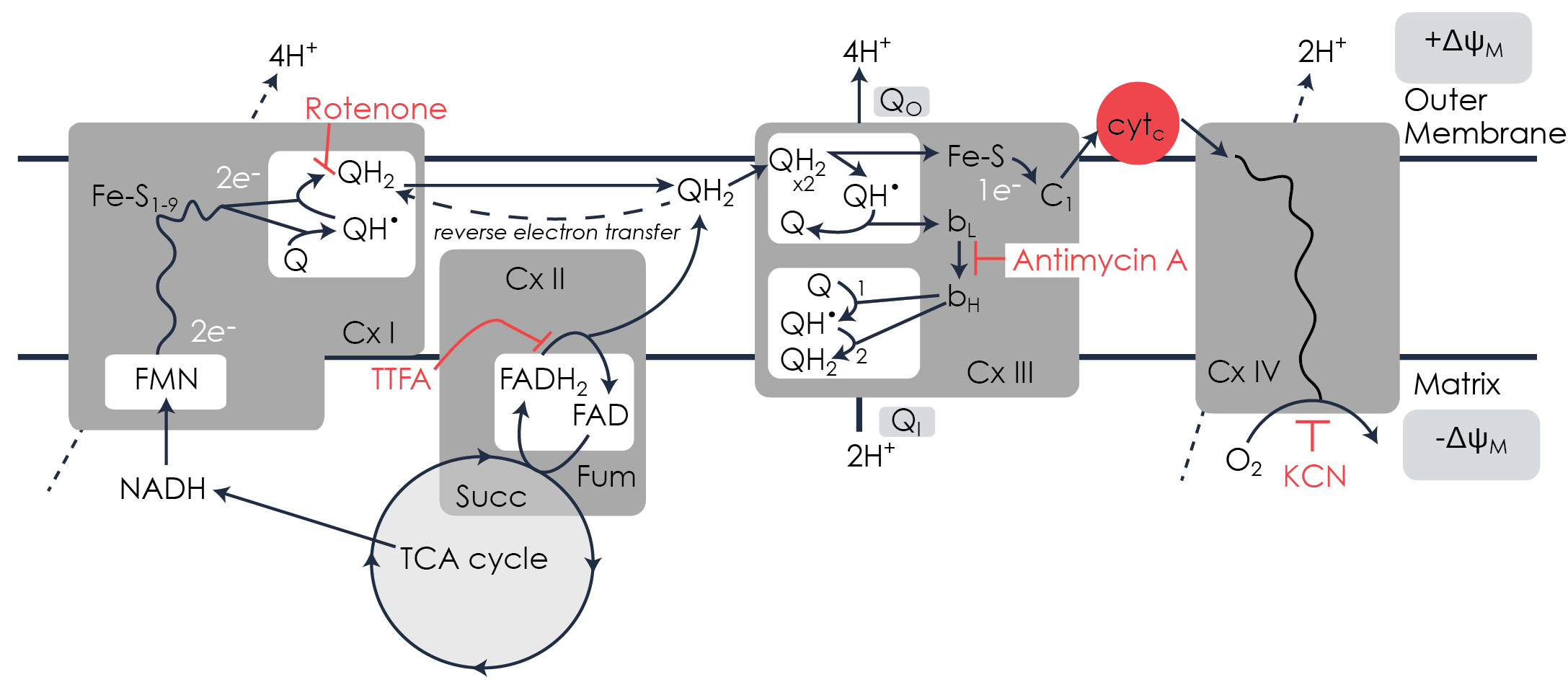
Figure 1. Enzymes and Inhibitors of the Electron Transport Chain
The generation of the proton gradient occurs through the utilization of the reducing equivalents NADH and FADH2, which are generated in the tricarboxylic acid cycle (TCA cycle). These reducing equivalents enter the ETC at Complex I (NADH dehydrogenase), which results in the translocation of protons (H+), and Complex II (succinate dehydrogenase), respectively. Following entry, electrons are shuttled from Complexes I and II via the lipophilic electron carrier ubiquinone (Q) to Complex III (ubiquinol cytochrome c oxidoreductase). Within Complex III, the redox cycling of ubiquinone, known as the Q-cycle, results in the translocation of protons. Ubiquinol primarily passes its electrons on to cytochrome cytc, however free radicals for cell signaling are also generated. Electrons that are transferred to cytochrome c are subsequently utilized in Complex IV (cytochrome c oxidase), leading to the step-wise reduction of O2 to H2O and resulting in the translocation of protons. 2-5
Given this unique and complex biochemistry, it is not surprising that mitochondria are often susceptible to drug-induced toxicity. Over the past two decades, 80% of the drugs pulled from the market or given black box warnings were flagged due to hepatotoxicity or cardiotoxicity complications resulting from mitochondrial toxicity.6,7 Furthermore, compounds that inhibit Complex I have been linked to neurological disorders such as Parkinson’s disease, while those that inhibit Complex II have been linked to increased tumorigenesis.8,9 A number of compounds exhibiting some degree of mitochondrial inhibition, including certain NSAIDs, antidepressants, drugs used to treat autoimmune disorders, and diabetes drugs have been approved by the FDA.10-15 Despite a presumed tolerable level, many of these drugs have the potential to disrupt the proton gradient and to decrease mitochondrial efficiency. Historically, drugs deemed toxic to mitochondria conspicuously induce substantial and life-threatening hepatotoxicity or cardiotoxicity. Mitochondrial toxicity has only recently been appreciated for its role in adverse drug reactions, thus prompting the pharmaceutical industry to implement mitochondrial toxicity screening platforms as a part of their ADME-Tox process. This mitochondrial toxicity screening, however, is not yet required by the FDA.
- Screen your compound for potential inhibition of the electron transport chain
- Designed as a colorimetric 96-well plate format; no special equipment required
- Isolated bovine heart mitochondria are provided in each kit
- Preincubation with antibodies is not required
- Amenable to high throughput screening
Given the complexity of the organelle, it is not surprising that there are multiple mechanisms by which a compound can impair mitochondrial function. Many of these mechanisms can be elucidated using prototypical mitochondrial inhibitors as probes. These classic inhibitors are complex specific, including rotenone (Complex I), 2-thenoyltrifluoracetone (TTFA) (Complex II), Antimycin A (Complex III), and potassium cyanide (KCN) (Complex IV). The sites at which these inhibitors bind are shown in Figure 1.
Despite the known adverse drug reactions associated with mitochondrial toxicity, few assays are available that allow for high throughput mitochondrial toxicity screening. Furthermore, the toxicity screening assays that are currently available employ expensive instrumentation and consumables, or require lengthy immunocapture-based reactions. Cayman has developed several MitoCheck Activity Assay Kits to screen for inhibitors of the mitochondrial ETC complexes. For end-user convenience, isolated bovine heart mitochondria are provided in each kit at optimal concentrations required for assaying activities of the individual complexes. These assays can be performed in a standard 96-well plate and do not require preincubation with antibodies, making them ideal for high throughput screening applications.
ETC Inhibition Analysis
Complex I activity was measured by monitoring the decrease in A340 corresponding to oxidation of NADH. Complex I assays were carried out in the presence of KCN (2 mM) to inhibit complex IV. Rotenone was titrated at 12-point half-log dilutions with a maximal concentration of 10 µM.
Complex II activity was measured as the succinate-dependent decrease in absorbance of DCPIP (A600). Complex II assays were carried out in the presence of rotenone (1 µM), antimycin A (10 µM), and KCN (2 mM) to inhibit Complexes I, III, and IV, respectively. TTFA was titrated in 12-point half log dilutions with a maximal concentration of 10 mM.
Complex II/III activity was measured as the succinate-dependent reduction of cytochrome c (A550). Complex II/III assays were carried out in the presence of rotenone (1 µM) and KCN (2 mM) to inhibit Complexes I and IV, respectively. Antimycin A was titrated at 12-point half-log concentrations with a maximal concentration of 10 µM.
Complex IV assays were carried out in the absence of mitochondrial inhibitors, using reduced cytochrome c as a substrate. Oxidation of cytochrome c was measured as a decrease in A550. KCN was titrated in 12-point half log dilutions with a maximal concentration of 2 mM.
Accurate IC 50s with a Time-Saving Advantage
The concentration response curves for inhibition of ETC complex I, II, III and IV generated using Cayman’s MitoCheck activity assays are shown in Figure 2. The calculated IC50 values for inhibition of complexes I-IV (Table 1) coincide with those formerly reported using previously developed immunocapture-based methods.16-19 Whereas these results are comparable to the cumbersome antibody-based assays, Cayman’s MitoCheck ETC Activity Assays offer an outstanding advantage in that ready-to-assay mitochondria are provided in the kits, eliminating that extra first step of isolation. Furthermore, lengthy preincubation with antibodies is not required. These time-saving features mean that a single assay to assess the mitochondrial toxicity of a drug candidate of interest can be completed in as little 30 minutes.
Figure 2. Concentration response curves of inhibitors of ETC complexes I, II, III and IV. See table below for calculated IC50 values obtained using Cayman's MitoCheck activity assay kits.
Figure 3. Inhibitor IC50s and assay variability obtained using Cayman Chemical's MitoCheck Activity Assays
References
1. Mitchell, P. and Moyle, J. Nature213, 137-139 (1967).
2. Trumpower, B. L. J. Biol. Chem.265, 11409-11412 (1990).
3. Chandel, N. S., McClintock, D. S., Feliciano, C. E., Wood, et al. J.Biol.Chem.275, 25130-25138 (2000).
4. Hoffman, D. L. and Brookes, P. S. J. Biol. Chem284, 16236-16245 (2009).
5. Campian, J. L., Gao, X., Qian, M., and Eaton, J. W. J. Biol. Chem.282, 12430-12438 (2007).
6. Dykens, J. A. and Will, Y. Drug discovery today12, 777-785 (2007).
7. Amacher, D. E. (2005). Curr. Med. Chem.12, 1829-1839 (2005).
8. Panov, A., Dikalov, S., Shalbuyeva, N., Taylor, et al. J. Biol. Chem.280, 42026-42035 (2005).
9. Dahia, P. L., Ross, K. N., Wright, M. E., Hayashida, C. Y. et al. PLoS Genet.1, 72-80 (2005).
10. Jacob, M., Bjarnason, I., and Simpson, R. J. Clin. Sci. (Lond) 101, 493-498 (2001).
11. Masubuchi, Y., Yamada, S., and Horie, T. Biochem. Pharmacol.58, 861-865 1999).
12. Bachmann, E. and Zbinden, G. Biochem. Pharmacol.28, 3519-3524 (1979).
13. Sprecher, H., Barr, H. M., Slotky, J. I., Tzukerman, M. et al. J. Biol. Chem.270, 20668-20676 (1995).
14. Yamamoto, N., Oliveira, M. B., Campello Ade, P., Lopes, L. C. et al. Cell. Biochem. Funct.6, 61-66 (1988).
15. Brunmair, B., Staniek, K., Gras, F., Scharf, N., et al. Diabetes.53, 1052-1059 (2004).
16. Nadanaciva, S., Bernal, A., Aggeler, R., Capaldi, R. et al. Toxicol. in vitro.21, 902-911 (2007).
17. Degli Esposti, M., Ghelli, A., Ratta, M., Cortes, D. et al. Biochem. J.301 ( Pt 1), 161-167 (1994)
18. Bougria, M., Vitorica, J., Cano, J., and Machado, A.. Eur. J. Pharmacol.291, 407-415 (1995)
19. Miyadera, H., Shiomi, K., Ui, H., Yamaguchi, Y. et al. P. Natl. Acad. Sci. USA.100, 473-477 (2003).
Contact Info
Cayman Chemical1180 East Ellsworth RoadAnn Arbor, Michigan 48108 USAToll Free: (800) 364-9897(USA and Canada only)Fax: (734) 971-3640


