
A convenient method for the direct visualization of S-glutathionylated proteins in whole (permeabilized) cells
Features
  • Direct visualization of S-glutathionylated proteins in whole (permeabilized) cells by flow cytometry and microscopy
  • Visualization using colorimetric or fluorescence detection
  • Reagents provided to test up to 30 samples
Technical Support & Resources

Visit our FAQ

Contact Us

Toll Free Phone (USA and Canada Only): (888) 526-5351
Direct Phone: (734) 975-3888

Request Technical Support

Technical Support Request

To streamline the process attach the appropriate questionnaire to your inquiry.

Download IHC QuestionnaireDownload WB Questionnaire

View Our Privacy Statement for details on how we use and protect your data. In addition, this site is protected by hCaptcha and its Privacy Policy and Terms of Service apply.

S-Glutathionylated Protein Detection Kit

Item No. 10010721
Technical Information
Synonyms
  • PSSG
Origin
Plant/Armoracia rusticana
Shipping & Storage Information
Storage
-20°C
Shipping
Wet ice in continental US; may vary elsewhere
Recommended Products

Certificates of Analysis & Batch Specific Data

Provide batch numbers separated by commas to download or request available product inserts, QC sheets, certificates of analysis, data packs, and GC-MS data.

    Add

    Bioanalysis & Assay Development Services
    Your Vision. Our Expertise.
    • Bioanalysis and compound screening using Cayman or qualified commercial assay kits
    • Development and qualification of fit-for-purpose assays in a variety of formats and platforms
    • Experienced scientists dedicated to developing the best strategy for your project
    LEARN MORE
    Product Description

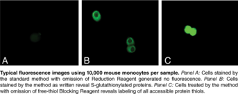
    Glutathione (GSH) is a tripeptide (γ-glutamylcysteinylglycine) widely distributed in both plants and animals.1,2 GSH is involved in maintenance of protein sulfhydryl reduction status.3,4 The concentration of GSH ranges from a few micromolar in plasma to several millimolar in tissues such as liver.5,6 Mixed protein glutathionyl disulfides are a post translational protein modification of growing interest.7,8,9 Protein S-glutathionylation (PSSG) may modify the activity of a large number of cell proteins, including metabolic, structural, cytoskeletal, and signaling proteins.10 PSSG detection methods can employ GSH adduct antibodies, GSH derivatives, and differential labeling systems based on the “Biotin-Switch” method.9,11,12 Cayman’s S-Glutathionylated Protein Detection Assay Kit provides a convenient method for the direct visualization of S-glutathionylated proteins in whole (permeabilized) cells by flow cytometry and microscopy as well as avidin overlay analysis.13 This cell-based assay starts with the modification of protein free-thiols groups followed by enzymatic cleavage of any protein-S-glutathione (PSSG) adducts present in the sample. Biotinylation of the newly-formed protein free-thiols provides the basis for visualization using streptavidin-based colorimetric or fluorescence detection. Reagents are provided to test three sets of 10 samples (most convenient) or up to thirty samples total at once if desired.

    WARNING This product is not for human or veterinary use.

    References & Product Citations
    Product Description References

    1. Foyer, C.H., Lelandais, M., and Kunert, K.J. Photooxidative stress in plants. Physiol. Plant. 92, 696-717 (1994).

    2. Bedia, C. Glutathione: Metabolism and function. (1976).

    3. Inoue, M., Saito, Y., Hirata, E., et alRegulation of redox states of plasma proteins by metabolism and transport of glutathione and related compounds. J. Protein Chem. 6, 207-225 (1987).

    4. Inoue, M. Interorgan metabolism and membrane transport of glutathione and related compounds. Renal Biochemistry 225-269 (1985).

    5. Lash, L.H., and Jones, D.P. Distribution of oxidized and reduced forms of glutathione and cysteine in rat plasma. Arch. Biochem. Biophys. 240, 583-592 (1985).

    6. Wendel, A., and Cikryt, P. The level and half-life of glutathione in human plasma. FEBS Lett. 120, 209-211 (1980).

    7. Pompella, A., Visvikis, A., Paolicchi, A., et alThe changing faces of glutathione, a cellular protagonist. Biochem. Pharmacol. 66(8), 1499-1503 (2003).

    8. Cotgreave, I.A. Analytical developments in the assay of intra-and extracellular GSH homeostsis: Specific protein S-glutathionylation, cellular GSH and mixed disulphide compartmentalisation and interstitial GSH redox balance. BioFactors 17, 269-277 (2003).

    9. Ying, J., Clavreul, N., Sethuraman, M., et alThiol oxidation in signaling and response to stress: Detection and quantification of physiological and pathophysiological thiol modifications. Free Radic. Biol. Med. 43, 1099-1108 (2007).

    10. Dalle-Donne, I., Rossi, R., Colombo, G., et alProtein S-glutathionylation: A regulatory device from bacteria to humans. Trends Biochem. Sci. 34(2), 85-96 (2008).

    11. Jaffrey, S.R., Erdjument-Bromage, H., Ferris, C.D., et alProtein S-nitrosylation: A physiological signal for neuronal nitric oxide. Nat. Cell Biol. 3(2), 193-197 (2001).

    12. Ckless, K., Reynaert, N.L., Taatjes, D.J., et alIn situ detection and visualization of S-nitrosylated proteins following chemical derivatization: Identification of Ran GTPase as a target for S-nitrosylation. Nitric Oxide 11, 216-227 (2004).

    13. Reynaert, N.L., Ckless, K., Guala, A.S., et alIn situ detection of S-glutathionylated proteins following glutaredoxin-1 catalyzed cysteine derivatization. Biochim. Biophys. Acta 1760, 380-387 (2006).

    Product Citations

    Marottoli, F.M., Trevino, T.N., Geng, X., et alAutocrine effects of brain endothelial cell-produced human apolipoprotein E on metabolism and inflammation in vitro. Front. Cell Dev. Biol. 9, (2021).

