
Technical Support & Resources
Visit our FAQ
Contact Us
Toll Free Phone (USA and Canada Only): (888) 526-5351
Direct Phone: (734) 975-3888
Product Categories
IκBα (Phospho-Ser32/36) Monoclonal Antibody (Clone 39A1413)
Item No. 13923
Technical Information
- I-kappa-B-alpha
- IkappaBalpha
- IKBA
- MAD-3
- MAD3
- Major Histocompatibility Complex Enhancer-binding Protein
- NF-kappa-B Inhibitor alpha
- NF-κB Inhibitor α
- NFKBIA
Shipping & Storage Information
Recommended Products
Certificates of Analysis & Batch Specific Data
Provide batch numbers separated by commas to download or request available product inserts, QC sheets, certificates of analysis, data packs, and GC-MS data.
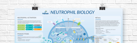
Neutrophil Biology Wall Poster
Explore how neutrophils shape the immune response in health and disease. This poster highlights neutrophil pathogen defense mechanisms, including phagocytosis, degranulation, and NETosis, as well as neutrophil roles in inflammation and NET-associated pathologies.
DOWNLOAD NOWProduct Description
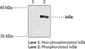
IκBα is an inhibitor of NF-κB and a member of the IκB family of proteins.1,2,3 It is composed of an N-terminal signal-receiving domain, which contains several post-translational modification sites that regulate its activity, an ankyrin-repeat domain, which contains six ankyrin repeats that mediate protein-protein interactions with NF-κB dimers, a proline-glutamic acid-serine-threonine (PEST) domain, and two nuclear export sequences.2 IκBα is ubiquitously expressed and shuttles between the cytoplasm and nucleus.1,2,3 IκBα is phosphorylated by the IκB kinase (IKK) complex, which is activated via downstream signaling events stimulated by TNF-α, other cytokines, viruses, or bacteria, and its phosphorylation leads to its ubiquitination and degradation by the proteasome. The degradation of IκBα results in unmasking of the nuclear localization signal of NF-κB dimers, which subsequently translocate to the nucleus and activate target genes. Mutations in NFKBIA, the gene encoding IκBα, are associated with anhidrotic ectodermal dysplasia with immunodeficiency.4 Cayman’s IκBα (Phospho-Ser32/36) Monoclonal Antibody (Clone 39A1413) can be used for immunoprecipitation (IP) and Western blot (WB) applications. The antibody recognizes IκBα (phospho-Ser32/36) from human, mouse, rat, porcine, bovine, and dog samples.
WARNING This product is not for human or veterinary use.
References & Product Citations
1. Regulation of IκBα function and NF-
2. Post-
3. Rel/NF-
4. Human IκBα gain of function: A severe and syndromic immunodeficiency. J. Clin. Immunol. 37(5), 397-412 (2017).
Contact Info
Cayman Chemical1180 East Ellsworth RoadAnn Arbor, Michigan 48108 USAToll Free: (800) 364-9897(USA and Canada only)Fax: (734) 971-3640


