
Technical Support & Resources
Visit our FAQ
Contact Us
Toll Free Phone (USA and Canada Only): (888) 526-5351
Direct Phone: (734) 975-3888
Product Categories
Product Type
Pertussis Toxin (islet-activating protein)
Item No. 19546
Technical Information
Shipping & Storage Information
Recommended Products
Certificates of Analysis & Batch Specific Data
Provide batch numbers separated by commas to download or request available product inserts, QC sheets, certificates of analysis, data packs, and GC-MS data.
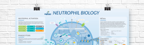
Neutrophil Biology Wall Poster
Explore how neutrophils shape the immune response in health and disease. This poster highlights neutrophil pathogen defense mechanisms, including phagocytosis, degranulation, and NETosis, as well as neutrophil roles in inflammation and NET-associated pathologies.
DOWNLOAD NOWProduct Description
Pertussis toxin (islet-activating protein) is a toxin, first isolated from B. pertussis, that is used to study G protein-coupled receptor signaling in cells and experimental autoimmune encephalomyelitis (EAE) in animals. Pertussis toxin catalyzes the transfer of the ADP-ribose moiety of NAD to the α subunits of heterotrimeric Gi/o proteins, resulting in the receptors being uncoupled from Gi/o proteins.1,2 Pertussis toxin is also used as an adjuvant, given with specific antigens, to immunize animals and induce EAE, an animal model of multiple sclerosis.3,4 Pertussis toxin was first described as an islet-activating protein because it caused a sustained potentiation of the secretory response of pancreatic islet cells to various stimuli that stimulate Gi-linked α-adrenergic receptors.5,6
WARNING This product is not for human or veterinary use.
References & Product Citations
1. Pertussis toxin and target eukaryotic cells: Binding, entry, and activation. The FASEB Journal 6(9), 2684-2690 (1992).
2. Islet-
3. Pertussis toxin modulates the immune response to neuroantigens injected in incomplete Freund’s adjuvant: Induction of Th1 cells and experimental autoimmune encephalomyelitis in the presence of high frequencies of Th2 cells. J. Immunol. 169(1), 117-125 (2002).
4. Experimental priming of encephalitogenic Th1/Th17 cells requires pertussis toxin-
5. The action of islet activating protein (pertussis toxin) on insulin’s ability to inhibit adenylate cyclase and activate cyclic AMP phosphodiesterases in hepatocytes. Biochem. J. 235(1), 145-149 (1986).
6. Slow interaction of islet-
Pharmacological inhibition of soluble epoxide hydrolase attenuates chronic experimental autoimmune encephalomyelitis by modulating inflammatory and anti-
Apoptotic caspases suppress type I interferon production via the cleavage of cGAS, MAVS, and IRF3. Mol. Cell 74(1), 19-31 (2019).
Contact Info
Cayman Chemical1180 East Ellsworth RoadAnn Arbor, Michigan 48108 USAToll Free: (800) 364-9897(USA and Canada only)Fax: (734) 971-3640


