
Technical Support & Resources
Visit our FAQ
Contact Us
Toll Free Phone (USA and Canada Only): (888) 526-5351
Direct Phone: (734) 975-3888
Product Categories
Research Area
Technical Information
- VIM
Shipping & Storage Information
Recommended Products
Certificates of Analysis & Batch Specific Data
Provide batch numbers separated by commas to download or request available product inserts, QC sheets, certificates of analysis, data packs, and GC-MS data.
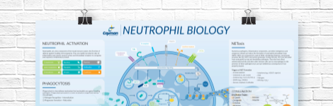
Neutrophil Biology Wall Poster
Explore how neutrophils shape the immune response in health and disease. This poster highlights neutrophil pathogen defense mechanisms, including phagocytosis, degranulation, and NETosis, as well as neutrophil roles in inflammation and NET-associated pathologies.
DOWNLOAD NOWProduct Description
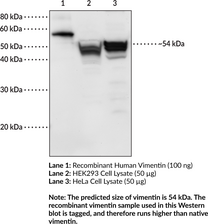
Vimentin is a cytoskeleton intermediate filament protein.1 It is composed of monomers that each contain a central α-helix rod domain, which facilitates formation of a coiled-coil dimer required for vimentin filament assembly, as well as N-terminal head and C-terminal tail domains.1,2 It is expressed in mesenchymal stem cells and cells of mesenchymal origin, including leukocytes, endothelial cells, and smooth muscle cells.3 Vimentin is attached to nuclei, endoplasmic reticulum, and mitochondria, and has a role in positioning organelles in the cytosol. It regulates glial morphology, facilitates motility and directional migration of fibroblasts, and is critical to mechanotransduction of shear stress and maintenance of vascular endothelial integrity.1 Vimentin controls transport of LDL-derived cholesterol from lysosomes to esterification sites.4 It is an aggresome component, forming a cage-like structure around aggregated, undegraded proteins at the microtubule organizing center.5 Vimentin is subject to citrullination under high calcium concentrations, which can occur during macrophage apoptosis, and citrullinated vimentin has been shown to have a role in the production of anti-citrullinated protein antibodies (ACPAs).6,7 ACPAs against citrullinated proteins, such as vimentin, are considered to be highly specific markers for rheumatoid arthritis and other autoimmune diseases.6 Cayman's Vimentin Monoclonal Antibody (Clone 12E4) can be used for ELISA and Western blot (WB) applications. The antibody recognizes vimentin at approximately 54 kDa from human samples.
WARNING This product is not for human or veterinary use.
References & Product Citations
1. Introducing intermediate filaments: From discovery to disease. J. Clin. Invest. 119(7), 1763-1771 (2009).
2. Assembling an intermediate filament network by dynamic cotranslation. J. Cell. Biol. 172(5), 747-758 (2006).
3. Formation of cytoskeletal elements during mouse embryogenesis. III. Primary mesenchymal cells and the first appearance of vimentin filaments. Differentiation 23(1), 43-59 (1982).
4. A functional role for vimentin intermediate filaments in the metabolism of lipoprotein-
5. Aggresomes: A cellular response to misfolded proteins. J. Cell Biol. 143(7), 1883-1898 (1998).
6. Clinical evaluation of anti-
7. Selective deimination of vimentin in calcium ionophore-
Contact Info
Cayman Chemical1180 East Ellsworth RoadAnn Arbor, Michigan 48108 USAToll Free: (800) 364-9897(USA and Canada only)Fax: (734) 971-3640


