
Technical Support & Resources
Visit our FAQ
Contact Us
Toll Free Phone (USA and Canada Only): (888) 526-5351
Direct Phone: (734) 975-3888
Product Categories
Technical Information
- CARD Adapter-Inducing Interferon-β
- Interferon-β Promoter Stimulator Protein 1
- Mitochondrial Antiviral-Signaling Protein
- Virus-Induced Signaling Adapter
Shipping & Storage Information
Recommended Products
Certificates of Analysis & Batch Specific Data
Provide batch numbers separated by commas to download or request available product inserts, QC sheets, certificates of analysis, data packs, and GC-MS data.
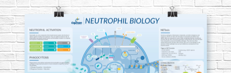
Neutrophil Biology Wall Poster
Explore how neutrophils shape the immune response in health and disease. This poster highlights neutrophil pathogen defense mechanisms, including phagocytosis, degranulation, and NETosis, as well as neutrophil roles in inflammation and NET-associated pathologies.
DOWNLOAD NOWProduct Description
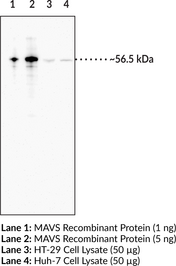
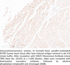
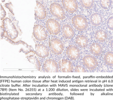
Mitochondrial antiviral-signaling protein (MAVS) is a mitochondrial membrane-bound adapter protein that initiates antiviral immunity.1 MAVS is activated upon viral infection by the dsRNA-sensing receptors RIG-I and MDA5 and by mitochondrial reactive oxygen species (ROS). Upon activation, MAVS polymerizes and binds to TANK-binding kinase 1 (TBK1) and IKKε, which form a signaling platform to activate IFN regulatory factor 3 (IRF3) and NF-ĸB, respectively.1,2 MAVS also mediates recruitment of the NLRP3 inflammasome to the mitochondria and activates caspase-1 to release IL-1β in response to non-crystalline inflammasome activators.3 Embryonic fibroblasts, peritoneal and bone marrow-derived macrophages (BMDMs), and conventional, but not plasmacytoid, dendritic cells from Mavs-/- mice exhibit reduced Sendai virus-induced increases in IFN-α and IFN-β secretion.4 Serum viral titers are increased and survival is decreased in Mavs-/- mice infected with vesicular stomatitis virus compared with wild-type controls. MAVS-deficient, SLE-prone Fcgr2b-/- mice have decreased serum levels of antinuclear autoantibodies and increased survival compared with MAVS-expressing Fcgr2b-/- mice.5 Aggregates of MAVS and plasma levels of IFN-β and the autoantibodies Sm and UN1RNP are increased in peripheral blood mononuclear cells (PBMCs) isolated from patients with systemic lupus erythematosus (SLE).6 Cayman's MAVS Monoclonal Antibody (Clone 7B9) can be used for ELISA, Immunohistochemistry, and Western blot applications. The antibody recognizes MAVS at 56.5 kDa from human samples.
WARNING This product is not for human or veterinary use.
References & Product Citations
1. Regulation of MAVS activation through post-
2. MAVS forms functional prion-
3. The adaptor MAVS promotes NLRP3 mitochondrial localization and inflammasome activation. Cell 153(2), 348-361 (2013).
4. The specific and essential role of MAVS in antiviral innate immune responses. Immunity 24(5), 633-642 (2006).
5. Antiviral adaptor MAVS promotes murine lupus with a B cell autonomous role. Front. Immunol. 10(2452), (2019).
6. Prion-
Contact Info
Cayman Chemical1180 East Ellsworth RoadAnn Arbor, Michigan 48108 USAToll Free: (800) 364-9897(USA and Canada only)Fax: (734) 971-3640


