
Technical Support & Resources
Visit our FAQ
Contact Us
Toll Free Phone (USA and Canada Only): (888) 526-5351
Direct Phone: (734) 975-3888
Technical Information
- MAP-2
- Microtubule-associated Protein 2
Shipping & Storage Information
Recommended Products
Certificates of Analysis & Batch Specific Data
Provide batch numbers separated by commas to download or request available product inserts, QC sheets, certificates of analysis, data packs, and GC-MS data.
Product Description
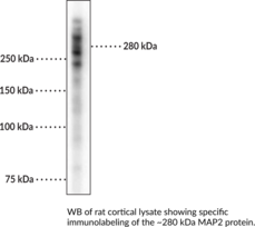
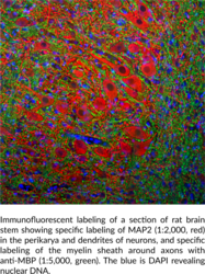
Microtubule-associated protein 2 (MAP2) belongs to the microtubule-associated protein (MAP) family of cytoskeletal filament proteins, which includes MAP1-5 and tau.1 MAP2 is composed of an N-terminal projection domain that interacts with PKA and a C-terminal domain that contains microtubule binding repeats containing a KXGS motif that is subject to phosphorylation.2 Five isoforms of MAP2 are generated through alternative splicing and are found in the cell body and dendrites of neurons, with some isoforms also found in astrocytes and oligodendrocytes of the CNS and in the testis.1,2 MAP2 is expressed in mature neurons, with the MAP2a and MAP2c isoforms expressed in a developmental stage-dependent manner while the MAP2b isoform is expressed throughout development and adulthood.1,3 MAP2 binds to and stabilizes microtubules and increases their rigidity, as well as interacts with F-actin during neurite initiation and with proteins involved in signal transduction.2 Phosphorylation of the KXGS motif by MAP/microtubule affinity-regulating kinase 1 (MARK1), MARK2, or PKA decreases the affinity of MAP2 for microtubules, and the detachment of MAP2 leads to microtubule destabilization and disassembly. Decreased levels of MAP2 protein have been found in postmortem brain tissue from patients with a Lewy body variant of Alzheimer’s disease.4 MAP2 has been found in cytoplasmic neuronal Lewy bodies colocalized with α-synuclein and ubiquitin in postmortem substantia nigra from patients with Parkinson’s disease.5 Cayman’s MAP2 (human) Monoclonal Antibody can be used for immunocytochemistry (ICC), immunohistochemistry (IHC), and Western blot (WB) applications. The antibody recognizes MAP2 at approximately 280 kDa from human, mouse, and rat samples.
WARNING This product is not for human or veterinary use.
References & Product Citations
1. .
2. .
3. .
4. .
5. .
Contact Info
Cayman Chemical1180 East Ellsworth RoadAnn Arbor, Michigan 48108 USAToll Free: (800) 364-9897(USA and Canada only)Fax: (734) 971-3640


