
Technical Support & Resources
Visit our FAQ
Contact Us
Toll Free Phone (USA and Canada Only): (888) 526-5351
Direct Phone: (734) 975-3888
Product Categories
Product Type
Application
Technical Information
- CSF-1
Shipping & Storage Information
Recommended Products
Certificates of Analysis & Batch Specific Data
Provide batch numbers separated by commas to download or request available product inserts, QC sheets, certificates of analysis, data packs, and GC-MS data.
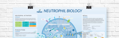
Neutrophil Biology Wall Poster
Explore how neutrophils shape the immune response in health and disease. This poster highlights neutrophil pathogen defense mechanisms, including phagocytosis, degranulation, and NETosis, as well as neutrophil roles in inflammation and NET-associated pathologies.
DOWNLOAD NOWProduct Description
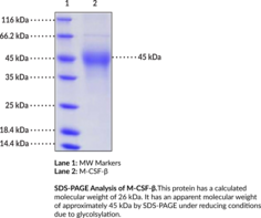
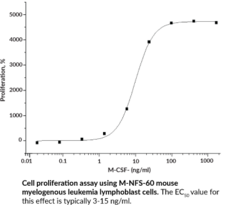
Macrophage colony-stimulating factor (M-CSF) is a glycoprotein encoded by Csf1 in mice that promotes the differentiation, proliferation, and function of mononuclear phagocytes, including macrophages, osteoclasts, and dendritic cells.1 Alternative splicing of Csf1 pre-mRNA produces one full-length long isoform (M-CSF-β), an intermediate-length isoform (M-CSF-γ), and a short-length isoform (M-CSF-α) that share sequence homology in the 150-amino acid receptor binding region which is required for the biological activity of M-CSF.2,3 M-CSF exists as a disulfide-linked homodimer where each monomer contains four α-helices, an anti-parallel β-sheet, and numerous glycosylation sites.1 M-CSF is constitutively produced by many cell types, including stromal cells, osteoclasts, fibroblasts, and macrophages, and is localized to the cell surface where it can be proteolytically cleaved to yield a secreted form.4,5 Binding of M-CSF to the M-CSF receptor, which is expressed by monocytes, macrophages, osteoclasts, and dendritic cells, promotes cell differentiation, proliferation, and survival of mononuclear phagocytes and regulates bone resorption by osteoclasts.6,4 Mice homozygous for Csf1op, an inactivating mutation, exhibit defects in fertility and neural development and develop osteopetrosis, a condition characterized by increased bone density.7 Neutralization of M-CSF with a monoclonal antibody decreases joint swelling and distortion in a mouse model of collagen-induced arthritis.8 M-CSF has been used to generate bone marrow- or peripheral blood monocyte-derived macrophages with an anti-inflammatory phenotype in vitro.9,10 Cayman's M-CSF-β (mouse, recombinant) protein can be used for binding and cell-based assay applications. This protein consists of 230 amino acids, has a calculated molecular weight of 26 kDa, and a predicted N-terminus of Lys33 after signal peptide cleavage. By SDS-PAGE, under reducing conditions, the observed molecular mass of the protein is approximately 45 kDa due to glycosylation.
WARNING This product is not for human or veterinary use.
References & Product Citations
1. Biology and action of colony-
2. Human macrophage-
3. M-
4. Macrophage colony-
5. Colony-
6. CSF-
7. Targeted disruption of the mouse colony-
8. The colony-
9. Biological role of granulocyte macrophage colony-
10. Toll-
Contact Info
Cayman Chemical1180 East Ellsworth RoadAnn Arbor, Michigan 48108 USAToll Free: (800) 364-9897(USA and Canada only)Fax: (734) 971-3640


