
Technical Support & Resources
Visit our FAQ
Contact Us
Toll Free Phone (USA and Canada Only): (888) 526-5351
Direct Phone: (734) 975-3888
SMAD1 (Phospho-Ser463,465)/SMAD5 (Phospho-Ser463,465)/SMAD9 (Phospho-Ser465,467
Item No. 42817
Technical Information
Shipping & Storage Information
Recommended Products
Certificates of Analysis & Batch Specific Data
Provide batch numbers separated by commas to download or request available product inserts, QC sheets, certificates of analysis, data packs, and GC-MS data.
Product Description
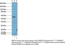
SMAD proteins are intracellular transcription factors and mediators of TGF-β signaling.1,2 They are composed of an N-terminal MAD homology 1 (MH1) domain containing nuclear localization and DNA-binding sequences and a C-terminal MH2 domain that is essential for SMAD-receptor interactions, as well as SMAD-SMAD interactions. Binding of TGF-β or bone morphogenetic protein (BMP) to their respective cell surface receptors initiates recruitment and activation of SMAD proteins. TGF-β induces SMAD2 and SMAD3 activation, while BMP binding induces activation of SMAD1, SMAD5, and SMAD9 via phosphorylation of serine 463 and 465 (Ser463,465) in SMAD1 and SMAD5 and Ser465,467 in SMAD9.1,2,3 SMAD1, -5, and -9 activation is associated with hypertrophy in patients with osteoarthritis, but associated with inhibition of fibrotic gene expression in various fibrotic diseases, including chronic kidney disease (CKD).3,2 Cayman’s SMAD1 (Phospho-Ser463,465)/SMAD5 (Phospho-Ser463,465)/SMAD9 (Phospho-Ser465,467) Rabbit Monoclonal Antibody (Clone RM487) can be used for Western blot (WB).
WARNING This product is not for human or veterinary use.
References & Product Citations
1. SMAD proteins: Mediators of diverse outcomes during infection. Eur. J. Cell Biol. 101(2), 151204 (2022).
2. New insights into TGF-
3. Separating friend from foe: Inhibition of TGF-
Contact Info
Cayman Chemical1180 East Ellsworth RoadAnn Arbor, Michigan 48108 USAToll Free: (800) 364-9897(USA and Canada only)Fax: (734) 971-3640


