
Technical Support & Resources
Information provided in the product description is from published literature. Due to the nature of scientific experimentation, your results (e.g., selectivity and effective concentrations) or specific application for this product may differ. If you have questions about how this product fits your application, please contact our technical support staff.
Visit our FAQ
Contact Us
Toll Free Phone (USA and Canada Only): (888) 526-5351
Direct Phone: (734) 975-3888
Product Categories
Technical Information
Shipping & Storage Information
Recommended Products
Certificates of Analysis & Batch Specific Data
Provide batch numbers separated by commas to download or request available product inserts, QC sheets, certificates of analysis, data packs, and GC-MS data.
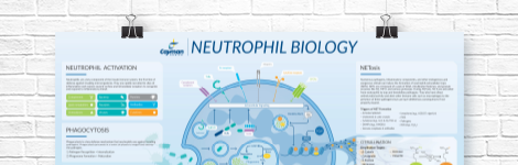
Neutrophil Biology Wall Poster
Explore how neutrophils shape the immune response in health and disease. This poster highlights neutrophil pathogen defense mechanisms, including phagocytosis, degranulation, and NETosis, as well as neutrophil roles in inflammation and NET-associated pathologies.
DOWNLOAD NOWProduct Description
Protonex™ Green 500-PEG12, SE is a PEGylated form of the amine-reactive and pH-dependent dye Protonex™ Green 500, SE with improved solubility that is intended for use in labeling proteins and antibodies. Protonex™ Green 500-PEG12, SE displays excitation/emission maxima of 453/522 nm, respectively, with high fluorescence intensity in acidic pH environments, such as phagosomes, endosomes, and lysosomes, but only weakly fluoresces in the extracellular environment. After conjugation, it can be used for microscopy and flow cytometry applications.
Unless otherwise noted, all unused stock solutions should be divided into single-use aliquots and stored at -20°C after preparation. Avoid repeated freeze-thaw cycles.
Protein stock solution (Solution A)
1. Mix 100 µl of a reaction buffer (e.g., 1 M sodium carbonate solution or 1 M phosphate buffer with pH ~9.0) with 900 µl of the target protein solution at a concentration of >2 mg/ml.
Note 1: The pH of the protein stock solution (Solution A) should be 8.5 ±0.5. If the pH of the protein solution is lower than 8.0, adjust the pH to the range of 8.0-9.0 using 1 M sodium bicarbonate solution or 1 M pH 9.0 phosphate buffer.
Note 2: The protein should be dissolved in 1X PBS, pH 7.2-7.4. If the protein is dissolved in Tris or glycine buffer, it should be dialyzed against 1X PBS to remove ammonium salts, such as ammonium sulfate and ammonium acetate, which are widely used for protein precipitation and may precipitate the protein, and free amines.
Note 3: Antibodies used for conjugation should be purified and free of sodium azide and thimerosal, which may interfere with the conjugation reaction. If sodium azide or thimerosal are present, use dialysis or a spin column to remove them. Conjugation of antibodies stabilized with BSA or gelatin will result in poor labeling.
Note 4: For optimal labeling efficiency, the recommended final protein concentration is 2-10 mg/ml.
Protonex™ Green 500-PEG12, SE stock solution (Solution B)
1. Add anhydrous DMSO into the vial of Protonex™ Green 500-PEG12, SE to make a 10 mM stock solution. Mix well by pipetting or vortex.
Note: Prepare the Protonex™ Green 500-PEG12, SE stock solution (Solution B) before starting the conjugation. Use promptly. Extended storage of the dye stock solution may reduce the dye activity. Solution B can be stored in the freezer for two weeks when protected from light and moisture. Avoid freeze-thaw cycles.
SAMPLE EXPERIMENTAL PROTOCOL
Important Note
This protocol was developed for conjugating Protonex™ Green 500-PEG12, SE to goat anti-mouse IgG. Additional optimization may be necessary for your specific protein.
Note: Each protein requires a distinct dye/protein ratio, which also depends on the properties of dyes. Over-labeling of a protein could detrimentally affect its binding affinity, while the protein conjugates of low dye/protein ratio give reduced sensitivity.
Run Conjugation Reaction
1. Use a 10:1 molar ratio of Solution B (dye)/Solution A (protein) as the starting point: Add 5 µl of the dye stock solution (Solution B, assuming the dye stock solution is 10 mM) into the vial of the protein solution (95 µl of Solution A) with effective shaking. The concentration of the protein is ~0.05 mM assuming the protein concentration is 10 mg/ml and the molecular weight of the protein is ~200 kDa.
Note: We recommend using a 10:1 molar ratio of Solution B (dye)/Solution A (protein). If it is too low or too high, determine the optimal dye/protein ratio at 5:1, 15:1, and 20:1, respectively.
2. Continue to rotate or shake the reaction mixture at room temperature for 30-60 minutes.
Purify the Conjugate
The following protocol is an example of dye-protein conjugate purification using a Sephadex G-25 column.
1. Prepare the Sephadex G-25 column according to the manufacturer's instructions.
2. Load the reaction mixture (from "Run conjugation reaction") to the top of the Sephadex G-25 column.
3. Add PBS (pH 7.2-7.4) as soon as the sample runs just below the top resin surface.
4. Add more PBS (pH 7.2-7.4) to the desired sample to complete the column purification. Combine the fractions that contain the desired dye-protein conjugate.
Note 1: For immediate use, the dye-protein conjugate should be diluted with staining buffer and aliquoted for multiple uses.
Note 2: For longer-term storage, the dye-protein conjugate solution should be concentrated or freeze-dried.
WARNING This product is not for human or veterinary use.
Contact Info
Cayman Chemical1180 East Ellsworth RoadAnn Arbor, Michigan 48108 USAToll Free: (800) 364-9897(USA and Canada only)Fax: (734) 971-3640


