
Features
- Homogeneous (no wash) TR-FRET assay method
- Rapid measurement of NF-κB protein levels in cells
- Streamlined “add-incubate-measure” assay protocol
- Contains TR-FRET antibody pairs, buffers, and positive control lysate
- Stable assay readout
- Compatible with both adherent and suspension cells
Technical Support & Resources
Visit our FAQ
Contact Us
Toll Free Phone (USA and Canada Only): (888) 526-5351
Direct Phone: (734) 975-3888
Product Categories
Product Type
Technical Information
- Nuclear Factor-κB
Shipping & Storage Information
Recommended Products
Certificates of Analysis & Batch Specific Data
Provide batch numbers separated by commas to download or request available product inserts, QC sheets, certificates of analysis, data packs, and GC-MS data.


Visit Our Cancer Resource Center
Find Tools & Resources to Study the Hallmarks of Cancer
- Cancer cell signaling & regulation
- Cancer metabolism
- Tumor microenvironment
Product Description
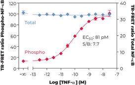
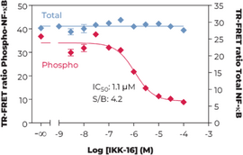
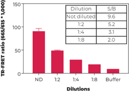
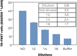
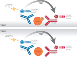
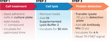
The Phospho-NF-kB (S536) + Total NF-kB assay kit is a homogeneous time-resolved Förster resonance energy transfer (TRFRET) sandwich immunoassay. The THUNDER™ Cell Signaling assay workflow consists of 3 steps. Following cell treatment, cells are first lysed with the specific Lysis Buffer provided in the kit. Then Phospho-NF-kB (S536) and Total NF-kB in the cell lysates are detected in separate wells with two pairs of fluorophore-labeled antibodies in a simple "add-incubate-measure" format (single-step reagent addition; no wash steps). For detection of the phosphorylated protein, one antibody is labeled with a donor fluorophore (Europium chelate; Eu-Ab1) and the second with a far-red acceptor fluorophore (FR-Ab2). The same approach is used for the second antibody pair detecting the total protein (Eu-Ab3 and FR-Ab4). The binding of the two matched labeled antibodies to distinct epitopes on the target protein (either phospho-NF-kB or total NF-kB) takes place in solution and brings the two dyes into close proximity. Excitation of the donor Europium chelate molecules with a flash lamp (320 or 340 nm) or a laser (337 nm) triggers a FRET from the donor to the acceptor molecules, which in turn emit a TR-FRET signal at 665 nm. Residual energy from the Eu chelate generates light at 615 nm. The signal at 665 nm is proportional to the concentration of Phospho-NF-kB (S536) and Total NF-kB in the cell lysate. Data can be expressed as either the signal at 665 nm or the 665 nm/615 nm ratio.
WARNING This product is not for human or veterinary use.
Contact Info
Cayman Chemical1180 East Ellsworth RoadAnn Arbor, Michigan 48108 USAToll Free: (800) 364-9897(USA and Canada only)Fax: (734) 971-3640


