
Features
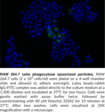
- Detect phagocytosis of fluorescein-labeled rabbit IgG-coated latex beads
- Validated in human peripheral blood monocytes and mouse peritoneal macrophages
- Phagocytosis detectable in 1-4 hours at 37°C
- Assess by flow cytometry or fluorescence microscopy
Technical Support & Resources
Visit our FAQ
Contact Us
Toll Free Phone (USA and Canada Only): (888) 526-5351
Direct Phone: (734) 975-3888
Product Categories
Product Type
Research Area
Application
Technical Information
Shipping & Storage Information
Recommended Products
Certificates of Analysis & Batch Specific Data
Provide batch numbers separated by commas to download or request available product inserts, QC sheets, certificates of analysis, data packs, and GC-MS data.
Bioanalysis & Assay Development Services
Your Vision. Our Expertise.
- Bioanalysis and compound screening using Cayman or qualified commercial assay kits
- Development and qualification of fit-for-purpose assays in a variety of formats and platforms
- Experienced scientists dedicated to developing the best strategy for your project
Product Description
Cayman’s Phagocytosis Assay Kit (IgG FITC) employs latex beads coated with fluorescently-labeled rabbit IgG as a probe for the measurement of the phagocytic process in vitro. The engulfed fluorescent beads can be detected using a fluorescence microscope, allowing kinetic studies of phagocytosis at the single-cell level. In addition, the flow cytometric readout provides the advantage of visualizing perturbations in phagocytosis on the population level and, when combined with antibody staining, of specific cell types within complex populations. This kit provides enough Latex Beads-Rabbit IgG-FITC Complex for up to 750 samples.
Needed but not supplied: Please download the kit booklet to verify if UltraPure Water (Milli-Q or equivalent) or any other components are needed for this assay.
WARNING This product is not for human or veterinary use.
References & Product Citations
Anti-
Paraventricular nucleus CRH neurons regulate acute lung injury via sympathetic nerve-
Macrophages derived from human induced pluripotent stem cells (iPSCs) serve as a high-
Atrazine induces phagocytotic dysfunction of microglia depends on nucleocytoplasmic translocation of acetylated HMGB1. Ecotoxicol. Environ. Saf. 252, 114583 (2023).
Tau activates microglia via the PQBP1-
FcγR engagement reprograms neutrophils into antigen cross-
Myeloperoxidase-
Contact Info
Cayman Chemical1180 East Ellsworth RoadAnn Arbor, Michigan 48108 USAToll Free: (800) 364-9897(USA and Canada only)Fax: (734) 971-3640


