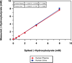
A sensitive tool for measuring β-HB levels in plasma, serum, or urine
Features
  • Measure β-HB from plasma, serum, and urine
  • Assay 40 samples in duplicate
  • Assay Range: 25-500 μM
  • Plate-based colorimetric measurement (445-455 nm)
Technical Support & Resources

Visit our FAQ

Contact Us

Toll Free Phone (USA and Canada Only): (888) 526-5351
Direct Phone: (734) 975-3888

Request Technical Support

Technical Support Request

To streamline the process attach the appropriate questionnaire to your inquiry.

Download IHC QuestionnaireDownload WB Questionnaire

View Our Privacy Statement for details on how we use and protect your data. In addition, this site is protected by hCaptcha and its Privacy Policy and Terms of Service apply.

β-Hydroxybutyrate (Ketone Body) Colorimetric Assay Kit

Item No. 700190

Technical Information
Synonyms
  • β-HB
  • 3-Hydroxybutyric Acid
Shipping & Storage Information
Storage
-20°C
Shipping
Wet ice in continental US; may vary elsewhere
Recommended Products

Certificates of Analysis & Batch Specific Data

Provide batch numbers separated by commas to download or request available product inserts, QC sheets, certificates of analysis, data packs, and GC-MS data.

    Add

    Lipid Resource Center
    Discover Products & Resources for Lipid Research
    • High-purity lipid standards
    • Lipid roles in biology
    • Lipids in health & disease
    • Lipids for pharmaceutical development
    • Protocols, advice, & resources
    EXPLORE NOW
    Product Description

    β-Hydroxybutyrate (β-HB; 3-hydroxybutyric acid) is a “ketone body” which is produced in the liver, mainly from the oxidation of fatty acids, and is exported to peripheral tissues for use as an energy source. The term ‘ketone body’ refers to three molecules, acetoacetate, β-HB, and acetone. β-HB and acetoacetate transport energy from the liver to the other tissues and acetone is generated by spontaneous decarboxylation of acetoacetate.1 The presence of ketosis may be normal or pathologic. Normally ketosis can indicate that lipid metabolism has been activated and the pathway of lipid degradation is intact. Normal ketosis is prevalent in many circumstances such as during fasting, after prolonged exercise or after a high fat diet. Pathological causes of ketosis include multiple organ failure, diabetes, childhood hypoglycemia, corticosteroid or growth hormone deficiency, intoxication with alcohol or salicylates and several inborn errors of metabolism.2 In acutely ill patients, these ketone bodies can accumulate in the body to cause ketoacidosis, which leads to the potentially life threatening condition known as metabolic acidosis.3 The presence and degree of ketosis can be determined by measuring blood levels of β-HB. Ordinarily, β-HB accounts for approximately 75% of the ketone bodies in serum.4,5,6 Measurement of β-HB provides a reliable index of the level of ketoacidosis, including the detection of subclinical ketosis.7,8,9 In diabetics, β-HB measurements (and blood glucose) can be used for the assessment of the severity of diabetic coma and is essential for the exclusion of hyperosmolar non-ketotic diabetic coma. The measurement of β-HB is also used to monitor insulin requirements, based on existing hyperketonemia.10 β-HB has more recently been evaluated for use in neurodegenerative diseases and inhibition of adipocyte lipolysis.11,12,13,14,15 Cayman’s β-HB (Ketone Body) Assay Kit provides a simple, reproducible, and sensitive tool for measuring β-HB levels in plasma, serum, urine, cell lysates, or tissue homogenates. The method for β-HB determination is based upon the oxidation of D-3-Hydroxybutyrate to acetoacetate by the enzyme 3-hydroxybutyrate dehydrogenase.16 Concomitant with this oxidation, the cofactor NAD+ is reduced to NADH. In the presence of diaphorase, NADH reacts with the colorimetric detector WST-1 to produce a formazan dye with an absorbance maximum at 445-455 nm. The absorbance of the dye is directly proportional to the β-HB concentration.

    Needed but not supplied: Please download the kit booklet to verify if UltraPure Water (Milli-Q or equivalent) or any other components are needed for this assay.

    WARNING This product is not for human or veterinary use.

    References & Product Citations
    Product Description References

    1. Guthrie, J.P., and Jordan, F. Amine-catalyzed decarboxylation of acetoacetic acid. The rate constant for decarboxylation of a β-imino acid. J. Am. Chem. Soc. 94(26), 9136-9141 (1972).

    2. Galán, A., Hernández, J.M., and Jimenez, O. Measurement of blood acetoacetate and β-hydroxybutyrate in an automatic analyser. J. Autom. Methods Manag. Chem. 23(3), 69-76 (2001).

    3. Foster, D.W., and McGarry, J.D. The metabolic derangements and treatment of diabetic ketoacidosis. N. Engl. J. Med. 309(3), 159-169 (1983).

    4. Persson, B. Determination of plasma acetoacetate and D-ß-hydroxy-butyrate in new-born infants by an enzymatic fluorometric micro-method. Scand. J. Clin. Lab. Invest. 25(1), 9-18 (1970).

    5. Wildenhoff, K.E. A micro-method for the enzymatic determination of acetoacetate and 3-hydroxybutyrate in blood and urine. Scand. J. Clin. Lab. Invest. 25(2), 171-179 (1970).

    6. Koch, D.D., and Feidbruegge, H. Optimized kinetic method for automated determination of β-hydroxybutyrate. Clin. Chem. 33(10), 1761-1766 (1987).

    7. Li, P.K., Lee, J.T., MacGillivray, M.H., et alDirect, fixed-time kinetic assays for β-hydroxybutyrate and acetoacetate with a centrifugal analyzer or a computer-backed spectrophotometer. Clin. Chem. 26(12), 1713-1717 (1980).

    8. Harano, Y., Kosugi, K., Hyosu, T., et alSensitive and simplified method for the differential determination of serum levels of ketone bodies. Clin. Chim. Acta 134, 327-336 (1983).

    9. MacGillivray, M.H., Li, P.K., Lee, J.T., et alElevated plasma ß-hydroxybutyrate concentrations without ketonuria in healthy insulin-dependent diabetic patients. J. Clin. Endocrinol. Metab. 54(3), 665-668 (1982).

    10. Alberti, K.G.M.M., and Hockaday, T.D.R. Rapid blood ketone body estimation in the diagnosis of diabetic ketoacidosis. Br. Med. J. 2, 565-568 (1972).

    11. Kashiwaya, Y., Takeshima, T., Mori, N., et alD-β-hydroxybutyrate protects neurons in models of Alzheimer’s and Parkinson’s disease. Proc. Natl. Acad. Sci. USA 97(10), 5440-5444 (2000).

    12. Tieu, K., Perier, C., Caspersen, C., et alD-β-hydroxybutyrate rescues mitochondrial respiration and mitigates features of Parkinson disease. J. Clin. Invest. 112(6), 892-901 (2003).

    13. Reger, M.A., Henderson, S.T., Hale, C., et alEffects of β-hydroxybutyrate on cognition in memory-impaired adults. Neurobiol. Aging 25, 311-314 (2004).

    14. Cheng, B., Yang, X., Hou, Z., et alD-β-hydroxybutyrate inhibits the apoptosis of PC12 cells induced by 6-OHDA in relation to up-regulating the ratio of Bcl-2/Bax mRNA. Auton. Neurosci. 134(1-2), 38-44 (2007).

    15. Taggart, A.K.P., Kero, J., Gan, X., et al(D)-β-hydroxybutyrate inhibits adipocyte lipolysis via the nicotinic acid receptor PUMA-G. The Journal of Biological Chemisty 280(29), 26649-26652 (2005).

    16. McMurray, C.H., Blanchflower, W.J., and Rice, D.A. Automated kinetic method for D-3-hydroxybutyrate in plasma or serum. Clin. Chem. 30(3), 421-425 (1984).

    Product Citations

    Patil, I., Sancheti, H., Stiles, B.L., et alBrain metabolic and functional alterations in a liver-specific PTEN knockout mouse model. PLoS One 13(9), e0204043 (2018).

    Harun-Or-Rashid, M., Pappenhagen, N., Palmer, P.G., et alStructural and functional rescue of chronic metabolically stressed optic nerves through respiration. J. Neurosci. 38(22), 5122-5139 (2018).

    Luo, W., Qin, L., Li, B., et alInactivation of HMGCL promotes proliferation and metastasis of nasopharyngeal carcinoma by suppressing oxidative stress. Sci. Rep. 7(1), 11954 (2017).

    Kephart, W.C., Mumford, P.W., Mao, X., et alThe 1-week and 8-month effects of a ketogenic diet or ketone salt supplementation on multi-organ markers of oxidative stress and mitochondrial function in rats. Nutrients 9(9), e1019 (2017).

    Wang, A., Huen, S.C., Luan, H.H., et alOpposing effects of fasting metabolism on tissue tolerance in bacterial and viral inflammation. Cell 166(6), 1512-1525 (2016).

    Geisler, C.E., Hepler, C., Higgins, M.R., et alHepatic adaptations to maintain metabolic homeostasis in response to fasting and refeeding in mice. Nutr. Metab. (Lond.) 13, 62 (2016).

    French, R.P., Lyle, J., Tracey, S., et alHigh survivorship after catch-and-release fishing suggests physiological resilience in the endothermic shortfin mako shark (Isurus oxyrinchus). Conserv. Physiol. 3(1), cov044 (2015).

    Allalou, A., Nalla, A., Prentice, K.J., et alA predictive metabolic signature for the transition from gestational diabetes mellitus to type 2 diabetes. Diabetes 65(9), 2529-2539 (2016).