
News & Announcements
Deoxysphingolipids
Article from 2018-08-10
Headless sphingolipids create toxic, dead-end metabolites
Sphingolipids are a broad class of lipids that play essential roles in cell membrane integrity, signal transmission, and cell recognition. Diversity in function of the different classes of sphingolipids (e.g., sphingomyelins, galactosylceramides, gangliosides) is built in with the addition of various functional headgroups, while the traditional backbone of most mammalian sphingolipids is consistent, typically a C18 (or C16) chain containing 2-amino, 1,3-diol moieties. Atypical sphingoid bases that lack the C1 hydroxyl group of regular sphingoid bases have now also been identified. These headless 1-deoxysphingoid bases are not able to undergo metabolism to form sphingomyelins, galactosylceramides, gangliosides, etc. and, instead, have been shown to cause cellular toxicity.
In mammals, sphingolipids are typically produced de novo by serine palmitoyltransferase (SPT), the rate-limiting enzyme that uses serine as its substrate (Figure 1). Atypical 1-deoxysphinganine and 1-desoxysphinganine formation results from reactions in which a mutated SPT utilizes alanine or glycine, respectively, instead of serine as a substrate (Figure 1). These atypical lipids have also been reported to be produced by the reaction of wild-type SPT with alanine or glycine.
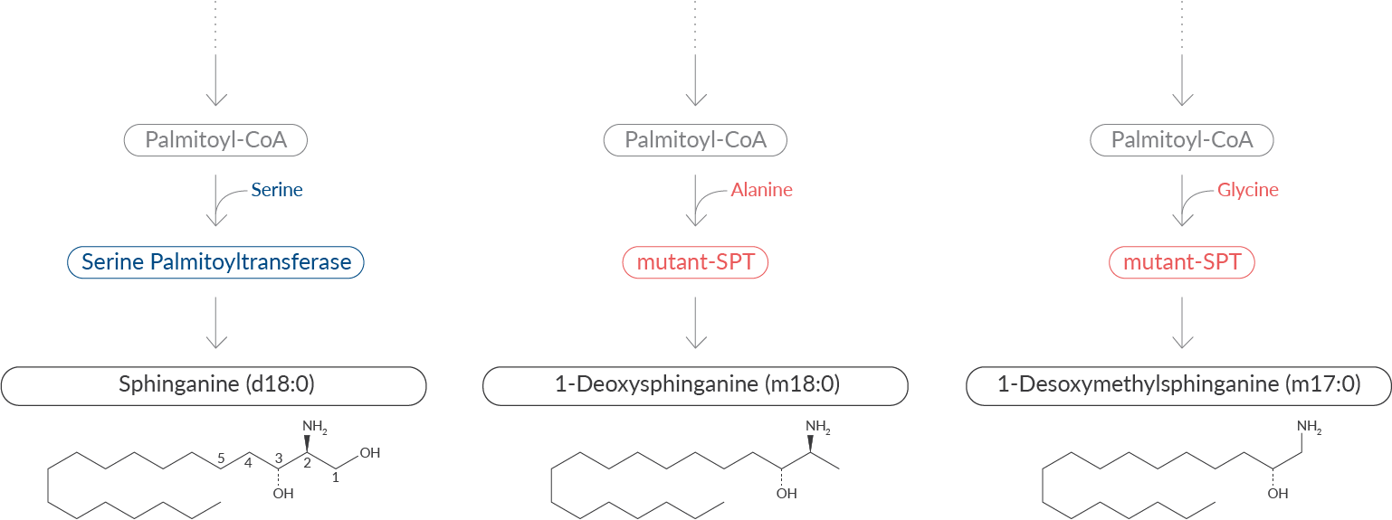
Figure 1. Sphingolipid biosynthesis via traditional (blue) and atypical (red) pathways.
Deoxysphinganines can be converted to 1-deoxysphingosines and 1-deoxyceramides. However, the missing OH-group prevents further metabolism to, sphingosine-1-phosphate (S1P). As a result, metabolism by S1P lyase, the canonical catabolic pathway, is lost, creating a dead-end to proper sphingosine degradation. These lipids possess highly toxic biophysical properties upon accumulation, and their metabolism, subcellular localization, and protein-lipid interactions are not well understood.
HSAN1
A rare genetic disorder, hereditary sensory and autonomic neuropathy type 1 (HSAN1) disease, is associated with cytotoxic accumulation of deoxysphingolipids. This disease is characterized by severe sensory loss (e.g., pain, temperature, and pressure) due to sensory axon degeneration and motor and autonomic neuron damage, which leads to injury, infection, muscle wasting, and motor impairment. HSAN1 results from multiple, different missense mutations in genes that code for the SPTLC1 and 2 subunits of SPT. These mutations induce a permanent shift in the substrate preference from serine to alanine, which results in the pathological formation of atypical and neurotoxic deoxysphingolipids. Studies in both mice bearing SPT mutations and a HSAN1 clinical trial have shown that serine supplementation can suppress formation of deoxysphingolipids and help to retain neurological function.
Metabolic Syndrome and Type 2 Diabetes
Deoxysphingolipids have also been shown to interfere with the survival of pancreatic β cells and insulin production. As such, plasma levels of deoxysphingolipids are higher in subjects with metabolic syndrome and type 2 diabetes and may serve as a predictive biomarker for these diseases. Deoxysphingolipid levels were also found elevated in plasma from patients exhibiting diabetic neuropathy. In common with animal models for HSAN1, serine supplementation has been used to successfully lower deoxysphingolipid levels in streptozotocin-induced diabetic rats, improving neuropathic hyperalgesia.
Mitochondrial Dysfunction and ER Stress
1-Deoxysphingolipids have been found to accumulate in mitochondria, causing swelling, fragmentation, and dysfunction. Morphological changes in the endoplasmic reticulum, including enlarged domains and increased density, have also been observed. Mitotoxicity and/or ER stress may underlie the neurotoxicity associated with HSAN1 and diabetic neuropathy.
Mycotoxin Analog
1-Deoxysphingolipids have been most extensively studied in the context of fumonisin B1, a fungal toxin that infests cattle feed, causing food poisoning. Structurally, fumonisin B1 appears as a deoxysphinganine analog, lacking the C1 hydroxyl group similar to that of other 1-deoxysphingoid bases. This mycotoxin disrupts sphingolipid metabolism by targeting ceramide synthase, the enzymes responsible for N-acylation of sphingolipids. Ceramide synthase inhibition results in the toxic accumulation of sphinganine, sphinganine-1-phosphate, etc. and suppresses ceramide synthesis, which can result in a wide spectrum of diseases in animals and humans. Fumonisin B1 is often used to as a tool to block ceramide production. It has also been used to rescue mitotoxicity in cultured fibroblasts exposed to 1-deoxysphinganine.
Native 1-Deoxysphingosine
Originally, the structure of 1-deoxysphingosine, as drawn on the left in Figure 2, was assumed to follow the canonical pathway of sphingolipid synthesis with a (4E) double bond in the n-14 position. In the traditional synthesis pathway, this bond would be introduced by sphingolipid Δ4-desaturase 1. Recently, however, the double bond in native 1-deoxysphingosine was found to be located at the n-4 (14Z) position (Figure 2).
Figure 2. The first synthesized 1-deoxysphingosine contained a (4E) double bond (left), based on assumptions of established sphingolipid biosynthesis. Recent discoveries, however, confirm that native 1-deoxysphingosine bears a (14Z) double bond (right).
This indicates that deoxysphingolipids are metabolized differently than traditional sphingolipids. Until recently, the native form of 1-deoxysphingosine was not commercially available to explore these differences. Cayman scientists have successfully synthesized 1-deoxysphingosine (m18:1(14Z)) to aid in a more detailed investigation of the native form of this lipid. A full line of 1-deoxysphinganine (m18:0) lipids and 1-deoxysphingosine (4E) lipids are also available in our product catalog. Indeed, much is left to be discovered in the enzymatic pathways, metabolic steps, and biological functions of these potentially toxic lipids.
1-Deoxysphinganine Line
C6 dihydro 1-Deoxyceramide (m18:0/6:0)
C22 dihydro 1-Deoxyceramide (m18:0/22:0)
C24 dihydro 1-Deoxyceramide (m18:0/24:0)
C24:1 dihydro 1-Deoxyceramide (m18:0/24:1)
1-Deoxysphingosine (14Z) Line
1-Deoxysphingosine (m18:1(14Z))
C6 1-Deoxyceramide (m18:1(14Z)/6:0)
C22 1-Deoxyceramide (m18:1(14Z)/22:0)
C24:1 1-Deoxyceramide (m18:1(14Z)/24:1)
1-Deoxysphingosine (4E) Line
1-Deoxysphingosine (m18:1(4E))
C6 1-Deoxyceramide (m18:1/6:0)
C12 1-Deoxyceramide (m18:1/12:0)
C16 1-Deoxyceramide (m18:1/16:0)
C22:1 1-Deoxyceramide (m18:1/22:1)
C24 1-Deoxyceramide (m18:1/24:0)
C24:1 1-Deoxyceramide (m18:1/24:1(15Z))
View complete details on 250+ sphingolipids available from Cayman
Need custom synthesis of a particular sphingolipid?
Email us at contractresearch@caymanchem.com
References
Alecu, I., Othman, A., Penno, A., et al. Cytotoxic 1-deoxysphingolipids are metabolized by a cytochrome P450-dependent pathway. J. Lipid Res.58(1), 60-71 (2017).
Alecu, I., Tedeschi, A., Behler, N., et al. Localization of 1-deoxysphingolipids to mitochondria induces mitochondrial dysfunction. J. Lipid Res. 58(1), 42-59 (2017).
Bode, H., Bourquin, F., Suriyanarayanan, S., et al. HSAN1 mutations in serine palmitoyltransferase reveal a close structure-function-phenotype relationship. Hum. Mol. Genet. 25(5), 853-865 (2016).
Dohrn, M.F., Othman, A., Hirshman, S.K., et al. Elevation of plasma 1-deoxy-sphingolipids in type 2 diabetes mellitus: A susceptibility to neuropathy? Eur. J. Neurol.22(5), 806-814, e55 (2015).
Duan, J. and Merrill, A.H., Jr. 1-Deoxysphingolipids encountered exogenously and made de novo: Dangerous mysteries inside an enigma. J. Biol. Chem. 290(25), 15380-15389 (2015).
Penno, A., Reilly, M.M., Houlden, H., et al. Hereditary sensory neuropathy type 1 is caused by the accumulation of two neurotoxic sphingolipids. J. Biol. Chem.285(15), 11178-11187 (2010).
Poad, B.L.J., Maccarone, A.T., Yu, H., et al. Differential-mobility spectrometry of 1-deoxysphingosine isomers: New insights into the gas phase structures of ionized lipids. Anal. Chem. 90(8), 5343-5351 (2018).
Steiner, R., Saied, E.M., Othman, A., et al. Elucidating the chemical structure of native 1-deoxysphingosine. J. Lipid Res.57(7), 1194-1203 (2016).
Zuellig, R.A., Hornemann, T., Othman, A., et al. Deoxysphingolipids, novel biomarkers for type 2 diabetes, are cytotoxic for insulin-producing cells. Diabetes 63(4), 1326-1339 (2014).
Contact Info
Cayman Chemical1180 East Ellsworth RoadAnn Arbor, Michigan 48108 USAToll Free: (800) 364-9897(USA and Canada only)Fax: (734) 971-3640


