
News & Announcements
Quantifying NETs
Article from 2018-11-06
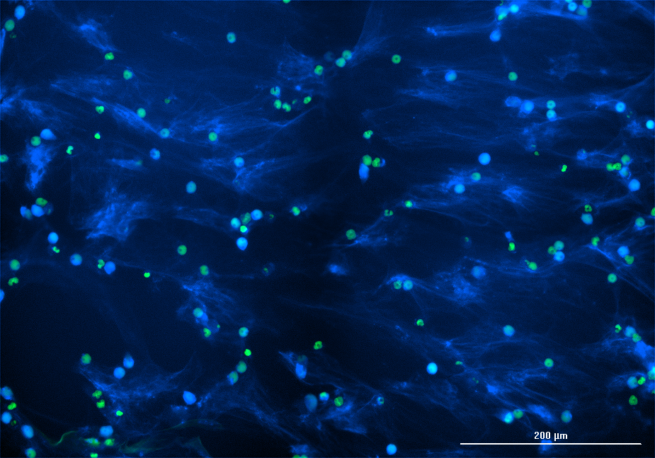
PMA induces NET formation in human neutrophils. Primary human neutrophils were stained with Nuclear Green DSC1 and stimulated with 20 nM PMA for 5 hours. Cells were fixed and stained with anti-MPO antibody followed by anti-mouse-DyLight™ 405. Nuclei are colored green and MPO is blue.
Neutrophil extracellular traps (NETs) are web-like structures extruded by neutrophils in response to infectious challenge. They are composed of DNA histones, and multiple antimicrobial agents (e.g., myeloperoxidase (MPO), elastase, defensins, etc.). Past attempts to detect and relatively quantify NETs have primarily relied on visualization by fluorescence microscopy (which is not accurately quantifiable) and/or measurement of extracellular DNA (which is not exclusive to NET formation). Most NET components are also released in soluble form from neutrophil granules (e.g., MPO, elastase, defensins) or during necrotic cell death (e.g., DNA and histones). Thus, these analytes are not NET-specific and cannot be relied on as accurate surrogates for measurement of NET formation. A handful of heterobifunctional ELISAs that detect various combinations of MPO, neutrophil elastase, and DNA have also been used, but these suffer from a lack of reliable, reproducible standards. To address these limitations, Cayman has developed the NETosis Assay Kit to study the process of NETosis induced ex vivo and the Citrullinated Histone H3 (Clone 11D3) ELISA Kit to accurately quantify an analyte produced almost exclusively during NET formation.
What is the difference between the NETosis Assay Kit and Citrullinated Histone H3 (Clone 11D3) ELISA Kit in terms of quantifying NETs?
Cayman's NETosis Assay Kit measures elastase unleashed from cultured primary neutrophils stimulated to release NETs with either PMA or a calcium ionophore (both are included in the kit). Notably, the kit does not depend upon the DNA component of NETs, as DNA release can occur independently of NETosis. As shown in the figure below, soluble (unbound) neutrophil elastase is washed away following NET generation. Upon digest of NET DNA by S7 nuclease, the supernatant containing NET-associated neutrophil elastase is added to a substrate, which is selectively cleaved by elastase to yield a 4-nitroaniline product that adsorbs light at 405 nm. The supernatant can also be tested for the presence of soluble dsDNA using a fluorescent DNA stain (e.g., PicoGreen).
Cayman’s Citrullinated Histone H3 (Clone 11D3) ELISA Kit quantifies the one NET component that is produced almost exclusively during NET formation. Histone H3 is citrullinated by peptidylarginine deiminase 4 (PAD4) at residues R2, R8, and R17 during the process of NET formation. In vitro, citrullinated H3 (CitH3) is released from neutrophils that have been stimulated with calcium ionophore during the process of NETosis and can be freed from NETs by treatment with nuclease. Thus, CitH3 could be considered a NET-specific biomarker. As shown in the image below, this ELISA is a sandwich assay that employs a capture monoclonal antibody directed against the citrullinated tail of histone H3 and a detection monoclonal antibody directed against a conserved epitope on the body of histone H3.
The NETosis Assay Kit detects elastase released from stimulated neutrophils. The Citrullinated Histone H3 ELISA measures CitH3, a NET-specific biomarker.
How was a suitable standard identified for the Citrullinated Histone H3 (Clone 11D3) ELISA Kit?
Histone H3 (human recombinant) was citrullinated by PAD4 (human recombinant) in a reaction buffer then the degree of citrullination was assessed using Cayman’s Citrulline-specific Probe and by mass spectrometry. Core Histones (human) isolated from HeLa cells, containing two copies of each of histones H2A, H2B, H3, and H4 were similarly citrullinated by PAD4 (human recombinant). Either Histone H3 (human recombinant) or Core Histones (human) function as an acceptable standard in the ELISA. The Citrullinated Histone H3 (Clone 11D3) ELISA Kit includes a proprietary formulation of CitH3 as the ELISA standard.
What stage of NETosis does the Citrullinated Histone H3 (Clone 11D3) ELISA Kit capture?
Human neutrophils release CitH3 into the supernatant within two hours of stimulation with a calcium ionophore (A23187), peaking at four hours as shown in the left graph below. Prior to two hours, CitH3 can be recovered from the detergent insoluble fraction of cell lysates, but not the supernatant. You can see NETosis early (while it is occurring) if you lyse the pre-NET cells in a detergent lysis buffer, centrifuge out the detergent insoluble fraction containing DNA and histones, and treat this with 15U/ml of S7 nuclease for 30 minutes until the pellet is dissolved. CitH3 will be in the detergent insoluble fraction of the cell lysate along with the DNA, so nuclease must release it before the ELISA can detect it.
Do medium supplements influence CitH3 production?
Cayman’s Citrullinated Histone H3 (Clone 11D3) ELISA Kit is recommened for use with RPMI-1640 basal medium supplemented with 10% FCS. As demonstrated in the graph below, this formulation has been shown to support CitH3 release from activated neutrophils. CitH3 cannot be detected if FCS is replaced with BSA or human albumin. This is because CitH3 is released from the neutrophil along with neutrophil elastase, whose function includes degrading histone tails, and serum contains alpha-1 antitrypsin, a natural inhibitor of elastase. Thus, in FCS, the elastase is inhibited, allowing the histone tails to remain intact for detection by the ELISA. Alpha-1 antitrypsin is not present in non-serum or non-plasma medium. Thus, the elastase can actively chew up histone tails, rendering them undetectable. Note that for this same reason, the buffer formulation recommended for Cayman’s NETosis Assay Kit is prepared using RPMI-1640 with 0.5% BSA—instead of serum—since the assay readouts elastase activity. Any alpha-1 antitrypsin present in serum would block this activity, so assay buffers from these two kits should not be shared.
The NETosis Assay Kit uses BSA in the assay buffer, while the Citrullinated Histone H3 ELISA requires FCS.
What species are compatible with these assays?
Cayman’s Citrullinated Histone H3 (Clone 11D3) ELISA Kit can detect CitH3 in cell culture supernatants, cell lysates, plasma, or serum from multiple species, including human and mouse. Additional species and sample types have not been tested and would need to be validated in your laboratory. Cayman’s NETosis Assay Kit can be used in any source of NET-producing cells. It is not possible to isolate large volumes of neutrophils from mouse blood, in which neutrophils comprise only ~10% of circulating white blood cells and collectible blood volumes are limited (~1 ml per mouse). Therefore, Cayman’s Neutrophil (mouse) Isolation Kit offers a streamlined method to isolate mouse neutrophils from peritoneal lavage or bone marrow. Some researchers have reported that it is difficult to generate NETs from mouse neutrophils obtained from bone marrow or peritoneal lavage. However, one published report has found that the addition of platelets to mouse neutrophils increases NET formation.1
1. Jiang, S., Park, D.W., Tadie, J.M., et al. Human resistin promotes neutrophil proinflammatory activation and neutrophil extracellular trap formation and increases severity of acute lung injury. J. Immunol. 192(10), 4795-4803 (2014).
The NETosis Assay Kit is an ex vivo neutrophil-based assay compatible with any species. The Citrullinated Histone H3 ELISA has been validated in mouse bone marrow leukocytes and serum as well as human plasma and serum.
What considerations should be made for blood sample collection and storage in terms of quantifying CitH3?
Serum and plasma contain nucleases and other factors that can interfere with the function of the Citrullinated Histone H3 (Clone 11D3) ELISA Kit. Serum or plasma prepared using citrate as the anticoagulant can be used without a purification step in the ELISA if they are first diluted a minimum of 1:2 in assay buffer. Plasma samples prepared with heparin or EDTA as the anticoagulant have been shown to cause slight interference in the assay with higher than expected recovery. Note that for Cayman’s NETosis Assay Kit, plasma and serum samples are not suitable sample types for this cell-based assay. Instead, neutrophils should be extracted from whole blood samples before proceeding with the assay.
Download our science poster to view the methods and data used to develop the CitH3 ELISA: Quantification of Neutrophil Extracellular Trap Formation Using an ELISA Specific for Citrullinated Histone H3.
You May Also Be Interested In
| | |

Contact Info
Cayman Chemical1180 East Ellsworth RoadAnn Arbor, Michigan 48108 USAToll Free: (800) 364-9897(USA and Canada only)Fax: (734) 971-3640


