
News & Announcements
Sphingomyelin and Lysosphingomyelin: Vital Membrane Components
Article from 2019-08-01
This article was originally published in the August 2019 edition of Matreya’s Newsletter for Glyco/Sphingolipid Research (PDF).
Sphingomyelin, dihydrosphingomyelin, and lysosphingomyelin (i.e., sphingosylphosphorylcholine) are major and important phosphosphingolipids found in mammalian cell membranes, especially in the membranes of the myelin sheath. Sphingomyelin is the most abundant sphingolipid in mammals and is found primarily in the exoplasmic leaflet of the membrane. Sphingomyelin has numerous critical cellular functions including signal transduction, apoptosis, and myelin sheath formation.1 Sphingomyelin also plays a significant role in Niemann-Pick disease (types A and B), multiple sclerosis, neonatal respiratory distress syndrome, and abetalipoproteinemia.
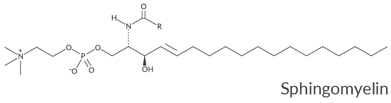
The ratio of sphingomyelin to ceramide in different cell types plays a critical role in cellular function.2 Sphingomyelin is an important amphiphilic component when plasma lipoprotein pools expand in response to large lipid loads or metabolic abnormalities.3 In contrast to ceramides, C6 sphingomyelin does not initiate vesicle formation in cells4 but has been used to enhance the uptake of antitumor drugs by cancer cells, thereby increasing their cytotoxicity.5 Lysosphingomyelin, the deacylated form of sphingomyelin, has been shown to induce intracellular calcium release while its short-chain analog, C2 sphingomyelin, requires a significantly higher concentration to initiate the same level of response.6
Sphingomyelin also has important implications in several severe diseases. Niemann-Pick disease is a rare lysosomal storage disorder with debilitating effects and is characterized by a deficiency of the enzyme acid sphingomyelinase. This results in the accumulation of sphingomyelin, leading to hepatosplenomegaly, liver dysfunction, interstitial lung disease, thrombocytopenia, anemia, an atherogenic lipid profile, bone disease, and neurodegeneration.7,8 Low levels of sphingomyelin are considered a blood biomarker for multiple sclerosis, although whether sphingomyelin is an active species in the disease remains unclear.9 In neonatal respiratory distress syndrome, the ratio of lecithin/sphingomyelin in amniotic fluid has been used to predict risk of the disease.10 An excess of sphingomyelin in red blood cells leads to abetalipoproteinemia, causing decreased membrane fluidity.11
Lysosphingomyelin has been identified in normal blood plasma, ascites, and various tissues. It is similar in structure to sphingosine-1-phosphate (S1P) and lysophosphatidylcholine (LPC) and has low-binding affinity to some of the same receptors, such as S1P receptors. It is a bioactive lipid that acts as an intracellular and extracellular signaling molecule in numerous biological processes such as vasoconstriction, vasodilation, angiogenesis, stress fiber formation, cytoskeletal rearrangements, proliferation, differentiation, migration, wound healing, and stimulation of DNA synthesis. Lysosphingomyelin can also inhibit the growth of various cell types, including tumor cells, causing much interest in its possible role as an antitumor therapy. It is a high-affinity ligand for the orphan receptor ovarian cancer G protein-coupled receptor 1 (OGR1). The specific binding of lysosphingomyelin to OGR1 also activates p42/44 mitogen-activated protein kinases (MAPKs) and inhibits cell proliferation.12 Lysosphingomyelin may be able to help treat inflammatory kidney diseases and has been found to activate various protein kinase cascades known to reduce inflammation. Lysosphingomyelin has also been shown to cause an increase in urine production in the kidneys with an abnormal accumulation of salt in the urine.13 Lysosphingomyelin acts as an inhibitor of calmodulin, a highly prevalent intracellular calcium sensor in eukaryotic cells.14
The extracellular effects of lysosphingomyelin appear to be stereospecific, while intracellular effects may not be. D-erythro-Lysosphingomyelin, but not L-threo-lysosphingomyelin, stereoselectively stimulates the proliferation of human adipose tissue-derived mesenchymal stem cells and stimulates an increase in calcium concentration and cellular proliferation.15 Both the L-threo-lysosphingomyelin isomer and the D-erythro-lysosphingomyelin isomer can act as second messengers by releasing calcium from internal stores.
Dihydrosphingomyelin, containing a saturated sphingosinebase, has been identified as a minor lipid component in many mammalian tissues but has recently been reported to be present in significant amounts in bovine brain and bovine milk.16 It is also found in much greater amounts in human lens membranes (half of all the phospholipids) where it has a critical role in ocular function and perhaps in age-related nuclear cataracts.17 However, dihydrosphingomyelin has been reported to be present only in small amounts in the lens membranes of other mammals. Dihydrosphingomyelin demonstrates good mixing properties with both sterols and sphingomyelin, indicating that it could function as a membrane organizer, and this may be the reason it is present in large amounts in human lens membranes where cholesterol is also enriched.18 The enzyme sphingomyelinase is active towards dihydrosphingomyelin and readily converts it to dihydroceramide. Dihydrosphingomyelin impairs HIV-1 infection by rigidifying liquid-ordered membrane domains, a finding that could have great potential in providing a therapeutic treatment for this debilitating disease.19
Available Sphingomyelins and Lysosphingomyelins
- Sphingomyelins (from bovine spinal cord)
- Sphingomyelins (buttermilk)
- Sphingomyelins (egg)
- Sphingomyelins (porcine RBC)
- C2 Sphingomyelin (d18:1/2:0)
- C16 Sphingomyelin (d18:1/16:0)
- C16 Sphingomyelin-d9 (d18:1/16:0-d9)
- C17 Sphingomyelin (d18:1/17:0)
- C17 D-erythro/L-threo Sphingomyelin (d18:1/17:0)
- C18 Sphingomyelin (d18:1/18:0)
- C20 Sphingomyelin (d18:1/20:0)
- C22 Sphingomyelin (d18:1/22:0)
- C16 Sphingomyelin-13C (d18:1/16:0-13C)
- C6 NBD Sphingomyelin (d18:1/6:0)
- C12 NBD Sphingomyelin (d18:1/12:0)
- D-erythro-Sphingosylphosphorylcholine exclusively available from Matreya
- L-threo Lysosphingomyelin (d18:1)
- D-erythro/L-threo Lysosphingomyelin (d18:1)
- Lysodihydrosphingomyelin (d18:0)
- Ceramide Phosphoethanolamines (bovine)
References
1. Kolesnick, R.N., Haimovitz-Friedman, A., and Fuks, Z. The sphingomyelin signal transduction pathway mediates apoptosis for tumor necrosis factor, Fas, and ionizing radiation. Biochem. Cell Biol. 72(11-12), 471-474 (1994).
2. Kilkus, J.P., Goswami, R., Dawson, S.A., et al. Differential regulation of sphingomyelin synthesis and catabolism in oligodendrocytes and neurons. J. Neurochem. 106(4), 1745–1757 (2008).
3. Nilsson, A. and Duan, R.D. Absorption and lipoprotein transport of sphingomyelin. J. Lipid Res. (47)1, 154-171 (2006).
4. Li, R., Blanchette-Mackie, J., and Ladisch, S. Induction of endocytic vesicles by exogenous C6-ceramide. J. Biol. Chem. 274(30), 21121-21127 (1999).
5. Veldman, R.J., Zerp, S., van Blitterswijk, W.J., et al. N-hexanoyl-sphingomyelin potentiates in vitro doxorubicin cytotoxicity by enhancing its cellular influx. Br. J. Cancer 90(4), 917-925 (2004).
6. Yule, D.I., Wu, D., Essington, T.E., et al. Sphingosine metabolism induces Ca2+ oscillations in rat pancreatic acinar cells. J. Biol. Chem. 268(17), 12353-12358 (1993).
7. Schmuth, M., Man, M.-Q., Weber, F., et al. Permeability barrier disorder in Niemann-Pick disease: Sphingomyelin-ceramide processing required for normal barrier homeostasis. J. Invest. Dermatol. 115(3) 459-466 (2000).
8. Cassiman, D., Packman, S., Bembi, B., et al. Cause of death in patients with chronic visceral and chronic neurovisceral acid sphingomyelinase deficiency (Niemann-Pick disease type B and B variant): Literature review and report of new cases. Mol. Genet. Metab. 118(3), 206-213 (2016).
9. Jacob, S., Al-Kandari, A., Alroughani, R., et al. Assessment of plasma biomarkers for their association with multiple sclerosis progression. J. Neuroimmunol. 305, 5-8 (2017).
10. St. Clair, C., Norwitz, E.R., Woensdregt, K., et al. The probability of neonatal respiratory distress syndrome as a function of gestational age and lecithin/sphingomyelin ratio. Am. J. Perinatol. 25(8), 473-480 (2008).
11. Barenholz, Y., Yechiel, E., Cohen, R., et al. Importance of cholesterol-phospholipid interaction in determining dynamics of normal and abetalipoproteinemia red blood cell membrane. Cell Biophys. 3(2), 115-126 (1981).
12. Xu, Y., Zhu, K., Hong, G., et al. Sphingosylphosphorylcholine is a ligand for ovarian cancer G-protein-coupled receptor 1. Nat. Cell Bio. 2(5), 261-267 (2000).
13. Xin, C., Ren, S., Eberhardt, W., et al. Sphingosylphosphorylcholine acts in an anti-inflammatory manner in renal mesangial cells by reducing interleukin-1β-induced prostaglandin E2 formation. J. Lipid Res. 48(9), 1985-1996 (2007).
14. Kovacs, E. and Liliom, K. Sphingosylphosphorylcholine as a novel calmodulin inhibitor. Biochem. J. 410(2), 427-437 (2008).
15. Jeon, E.S., Song, H.Y., Kim, M.R., et al. Sphingosylphosphorylcholine induces proliferation of human adipose tissue-derived mesenchymal stem cells via activation of JNK. J. Lipid Res. 47(3), 653-664 (2006).
16. Byrdwell, W.C. and Perry, R.H. Liquid chromatography with dual parallel mass spectrometry and 31P nuclear magnetic resonance spectroscopy for analysis of sphingomyelin and dihydrosphingomyelin II. Bovine milk sphingolipids. J. Chromatogr. A. 1146(2), 164-185 (2007).
17. Deeley, J.M., Hankin, J.A., Friedrich, M.G., et al. Sphingolipid distribution changes with age in the human lens. J. Lipid Res. 51(9), 2753-2760 (2010).
18. Nyholm, T.K., Nylund, M., and Slotte, J.P. A calorimetric study of binary mixtures of dihydrosphingomyelin and sterols, sphingomyelin, or phosphatidylcholine. Biophys. J. 84(5), 3138-3146 (2003).
19. Vieira, C.R., Munoz-Olaya, J.M., Sot, J., et al. Dihydrosphingomyelin impairs HIV-1 infection by rigidifying liquid-ordered membrane domains. Chem. Biol. 17(7), 766-775 (2010).
Contact Info
Cayman Chemical1180 East Ellsworth RoadAnn Arbor, Michigan 48108 USAToll Free: (800) 364-9897(USA and Canada only)Fax: (734) 971-3640


