
News & Announcements
Visualizing NETosis Kinetics
Article from 2019-02-26
Two-color, live-cell, image-based assay enables visualization plus quantification of NETosis kinetics
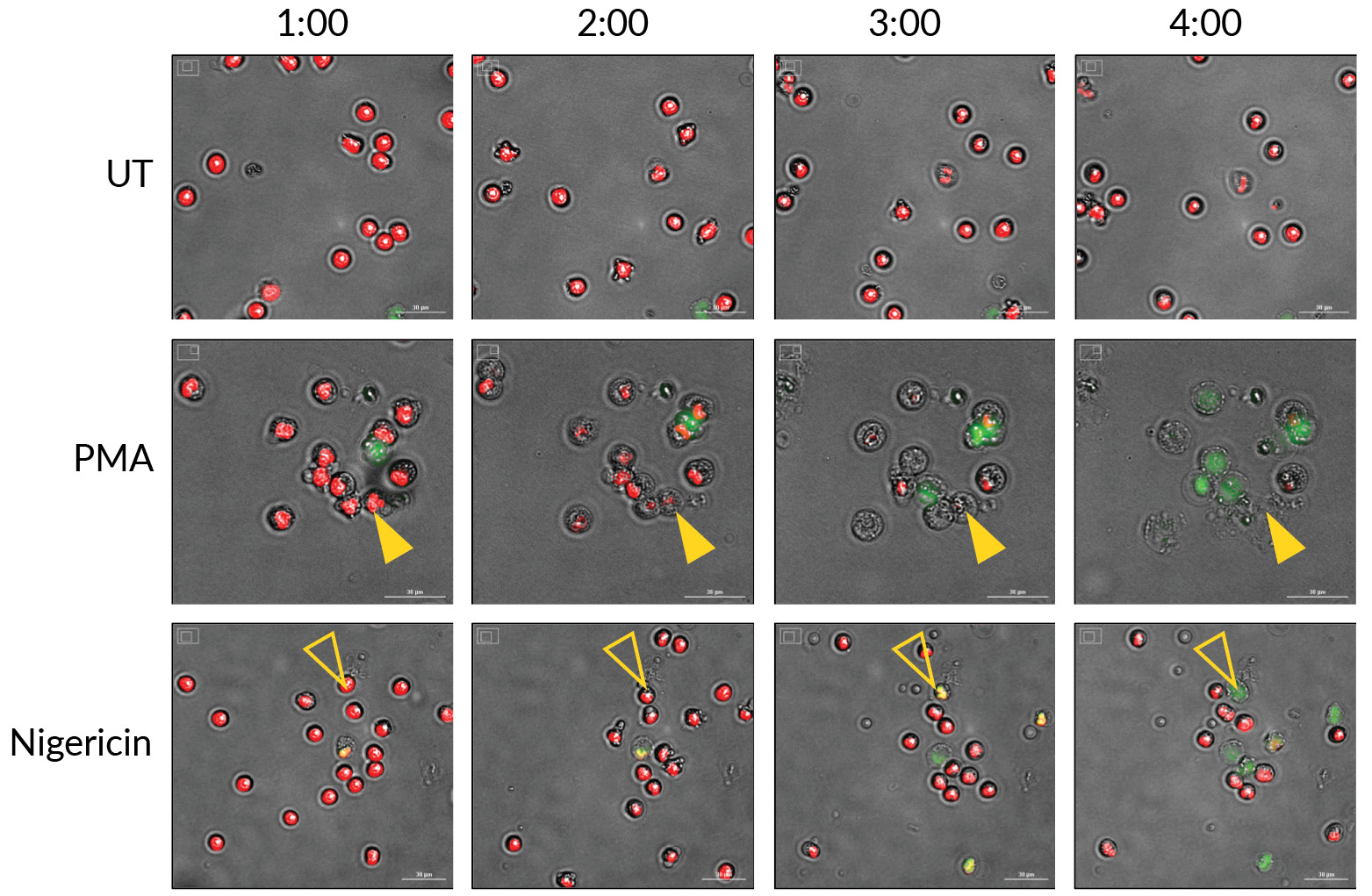
Monitoring the morphology of primary human neutrophils undergoing NETosis. DNA extruded during the dynamic process of NETosis is detected by Extracellular Nuclear Green™ Reagent in neutrophils stimulated with phorbol 12-myristate 13-acetate (PMA) or nigericin. Intact nuclei are counterstained with Permeable Nuclear Red™ Reagent. Timed images (every hour) were generated with BioTek®’s Cytation™ 5 Cell Imaging Multi-Mode Reader using a 20X objective and GFP and Cy5 LED filter combinations with brightfield. Filled and empty arrowheads follow individual cells undergoing NETosis. Scale bars are 30 μm.
As an important component of the innate immune system, neutrophil extracellular traps (NETs) can trap and immobilize pathogens, alert other immune cells such as macrophages to clear these pathogens, and have direct antimicrobial activity. The process by which NETs are released is a recently described, distinct form of programmed cell death called NETosis. Its mechanisms of action still require a deeper understanding since deviations in NETosis or defects in NET clearance have been implicated in a host of inflammatory disorders and autoimmune diseases, including systemic lupus erythematosus, psoriasis, and rheumatoid arthritis. Most attempts to study NETs have primarily relied on visualization by fluorescence microscopy and/or measurement of extracellular DNA, though both of these approaches have known limitations. Cayman has developed an easy staining protocol for visualizing the process of NETosis kinetically ex vivo and in vitro as an improvement over the traditional methods for NETosis analyses that are currently available.
Cayman’s NETosis Imaging Assay Kit utilizes the membrane permeability properties of two fluorescent DNA dyes—one cell permeable and one impermeable—to enable the visualization of NETosis dynamics over time. These dyes are provided in amounts sufficient for two 96-well plates. Characteristics of these dyes are as follows:
| Dye | Excitation (nm) | Emission (nm) | Typical Filter | Cell permeable? | What it stains |
|---|---|---|---|---|---|
| Extracellular Nuclear GreenTM Reagent (5 mM) | 503 | 526 | GFP/FITC | no | Extracellular DNA/permeabilized cells |
| Permeable Nuclear RedTM Reagent (5 mM) | 622 | 645 | Cy5 | yes | All nuclei |
To perform the assay, isolated neutrophils are stained with Nuclear Red™ for 15-30 minutes, then stained with Nuclear Green™ and stimulated to induce NETosis. Using brightfield, GFP, and Cy5 filter sets, images can be captured every 30 minutes to monitor NETosis kinetics while the plate is maintained at 37°C. Nuclear Red™ will mark the nuclei of every neutrophil. Any loss of nuclear lobulation can be visualized under brightfield. For neutrophils undergoing NETosis, chromatin decondensation is detected as an increase in nuclear diameter and a decrease in the intensity of Nuclear Red™ fluorescence. As DNA content mixes with cytoplasmic content and the cell membrane eventually becomes permeable, nuclear content will be stained with Nuclear Green™ present in the media. The release of NETs to the extracellular space is monitored by the increase of Nuclear Green™ fluorescence.
NETs are composed of DNA, histones, neutrophil elastase, myeloperoxidase, and other microbicidal proteins. Why use both intracellular and extracellular DNA detection to visualize NETosis kinetics?
Although neither DNA nor neutrophil elastase is exclusively released by neutrophils undergoing NETosis, traditional analyses of NETosis have relied on the quantification of these NET components. Unfortunately, these established methods typically rely on the visualization of fixed cells, which only capture a brief window into the process of NETosis. Since the process of NETosis occurs over a period of hours, quantifying such parameters at a single time point is limiting and makes the kinetics of NETosis challenging to study. Cayman’s simple staining protocol is designed to study the process of NETosis as it occurs by observing living neutrophils with a high-content imaging system.
The cell-permeable DNA dye (Nuclear RedTM) included in this kit allows visualization of the dynamic changes of the nucleus during the process of NETosis, while the cell-impermeable dye (Nuclear GreenTM) detects the extruded DNA. By combining the fluorescence visualization of these dyes with brightfield imaging in a high-content platform, different modes of cell death can be differentiated based on distinct changes in nuclear morphology and membrane integrity. This is important because differentiating between NETosis or apoptosis and necrosis can be informative about the stimulus used or experimental variables studied. Importantly, neutrophils may expel NETs without inducing cell lysis. In this case, the neutrophil becomes a cytoplast or “neutrophil ghost” whose chemotactic and phagocytic functions remain intact. This membrane integrity-dependent, dual-dye, live-content imaging assay platform provides a powerful tool to assess both neutrophil physiology and NETosis and can be used to develop and test novel neutrophil inhibitors.
Can the kit be used with a conventional microscope instead of a high-content imaging platform?
High-content imaging platforms allow for the percentage of neutrophils undergoing NETosis to be quantified kinetically, using any stimulus. The dose-dependent effect of inhibitors of NETosis can also be quantified to enable the rapid development of novel neutrophil targets. Using such a platform allows for a rapid, unbiased, reproducible, and more sensitive means to observe the kinetics of NETosis compared to conventional fluorescence microscopy.
What species/sample types are compatible with this assay?
This kit can be used in any source of NET-producing cells. The kit contains a ready-to-use Histopaque® reagent and protocol for isolating human peripheral blood neutrophils through centrifugation.
Note that for mouse studies, neutrophils typically comprise less than 10% of circulating white blood cells and collectible blood volumes are limited (~1 ml per mouse), so often it is not possible to isolate sufficient cells for study. As a solution to this issue, Cayman’s Neutrophil (mouse) Isolation Kit offers a streamlined method to isolate mouse neutrophils from peritoneal lavage or bone marrow. Some researchers have reported that it is difficult to generate NETs from mouse neutrophils obtained from bone marrow or peritoneal lavage. However, a published report has found that the addition of platelets to mouse neutrophils increases NET formation.1
1. Etulain, J., Martinod, K., Wong, S.L., et al. P-selectin promotes neutrophil extracellular trap formation in mice. Blood 126(2), 242-246 (2015).
What stimuli can be used to induce NETosis?
Neutrophils undergoing NETosis induced by various physiological stimuli show distinct changes, with a loss of multilobulated nuclei and nuclear decondensation, leading to membrane compromise and extrusion of DNA. The assay kit includes PMA and a calcium ionophore (A23187) as positive controls to stimulate neutrophils to produce NETs. Many other stimuli can be used as well, from other ionophores like the bacterial toxin nigericin and cellular products like resistin2 to opsonized pathogens like Candida albicans or Group B Streptococcus.3 Pathways to NETosis can also be dissected by combining chemical stimulators of NETosis with specific inhibitors of cellular processes required for NETosis.
2. Jiang, S., Park, D.W., Tadie, J.-M., et al. Human resistin promotes neutrophil proinflammatory activation and neutrophil extracellular trap formation and increases severity of acute lung injury. J. Immunol. 192(10), 4795-4803 (2014).
3. Kenny, E.F., Herzig, A., Krüger, R. et al., Diverse stimuli engage different neutrophil extracellular trap pathways. eLife 6:e24437 (2017).
Watch NET formation with time-lapse imaging of neutrophils.
Primary human neutrophils were stained with Permeable Nuclear Red™ Reagent and Extracellular Nuclear Green™ Reagent and stimulated with 20 nM PMA. Images were taken every 30 minutes with BioTek’s Cytation™ 5 Cell Imaging Multi-Mode Reader using brightfield, GFP, and Cy5 LED/filter sets. All nuclei are red, while only nuclei of membrane-compromised cells are green. Neutrophil NETs are visible as indistinct clouds of green, which form as distinct red nuclei, fade, and are extruded.
Read our article on Quantifying NETs to learn more about Cayman’s suite of NETosis assays
You May Also Be Interested In
| | |

Contact Info
Cayman Chemical1180 East Ellsworth RoadAnn Arbor, Michigan 48108 USAToll Free: (800) 364-9897(USA and Canada only)Fax: (734) 971-3640


