
Technical Support & Resources
Visit our FAQ
Contact Us
Toll Free Phone (USA and Canada Only): (888) 526-5351
Direct Phone: (734) 975-3888
Product Categories
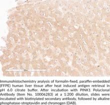
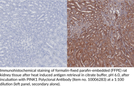
Technical Information
- BRPK
- PARK6
- PTEN Induced Putative Kinase 1
- PTEN Inducible Kinase 1
- Serine/Threonine-protein Kinase PINK1
Shipping & Storage Information
Recommended Products
Certificates of Analysis & Batch Specific Data
Provide batch numbers separated by commas to download or request available product inserts, QC sheets, certificates of analysis, data packs, and GC-MS data.
Product Description
PTEN-induced putative kinase 1 (PINK1) is a serine/threonine protein kinase that has a role in mitochondrial function.1,2 It is comprised of an N-terminal mitochondrial targeting sequence, a transmembrane domain, a serine/threonine kinase domain, and a C-terminal region.2 PINK1 is ubiquitously expressed primarily in the brain, skeletal muscle, and heart.3 It localizes to the mitochondria where it is either rapidly degraded or, under conditions of low mitochondrial membrane potential, accumulates on the outer mitochondrial membrane, where it recruits and activates the cytosolic E3 ubiquitin ligase Parkin, which targets the mitochondria for mitophagy.3,1 Pink1 knockout in rats leads to an age-dependent loss of dopaminergic neurons in the substantia nigra, as well as deficits in motor function and mitochondrial respiration.4 In mice, Pink1 knockout does not induce a loss of dopaminergic neurons without concomitant overexpression of α-synuclein in the substantia nigra.5 Loss-of-function mutations in PINK1 are causally associated with autosomal recessive early-onset Parkinson’s disease.3,6 Cayman’s PINK1 Polyclonal Antibody can be used for immunohistochemistry (IHC) and Western blot (WB) applications. The antibody primarily recognizes full-length PINK1 at approximately 66 kDa from human, mouse, and rat samples but also detects a truncated form of the protein at approximately 33 kDa.
WARNING This product is not for human or veterinary use.
References & Product Citations
1. Structure and function of parkin, PINK1, and DJ-
2. Analysis of the regulatory and catalytic domains of PTEN-
3. Parkin and PINK1 functions in oxidative stress and neurodegeneration. Brain Res. Bull. 133, 51-59 (2017).
4. New developments in genetic rat models of Parkinson’s disease. Mov. Disord. 33(5), 717-729 (2018).
5. Alpha-
6. Hereditary early-
PINK1 is dispensable for mitochondrial recruitment of parkin and activation of mitophagy in cardiac myocytes. PLoS One 10(6), e010707 (2015).
Contact Info
Cayman Chemical1180 East Ellsworth RoadAnn Arbor, Michigan 48108 USAToll Free: (800) 364-9897(USA and Canada only)Fax: (734) 971-3640


