
Features
- Measure mitochondrial membrane potential as an indicator of cell health
- Utilizes JC-1, a fluorescent, lipophilic, cationic dye
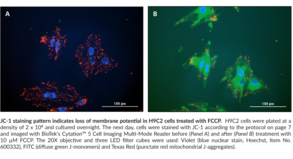
- Red fluorescence indicates healthy mitochondria and green fluorescence indicates mitochondria in poor health
- Optimized for fluorescence microscopy or fluorescent plate readers. An assay optimized for flow cytometry is available under Item No. 701560.
Technical Support & Resources
Visit our FAQ
Contact Us
Toll Free Phone (USA and Canada Only): (888) 526-5351
Direct Phone: (734) 975-3888
Product Categories
Product Type
Application
Technical Information
Shipping & Storage Information
Recommended Products
Certificates of Analysis & Batch Specific Data
Provide batch numbers separated by commas to download or request available product inserts, QC sheets, certificates of analysis, data packs, and GC-MS data.

Visit Our Cancer Resource Center
Find Tools & Resources to Study the Hallmarks of Cancer
- Cancer cell signaling & regulation
- Cancer metabolism
- Tumor microenvironment
Product Description
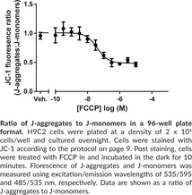
Mitochondrial membrane potential, (ΔψM), is an important parameter of mitochondrial function used as an indicator of cell health. JC-1 is a lipophilic, cationic dye that can selectively enter into mitochondria and reversibly change color from green to red as the membrane potential increases. In healthy cells with high mitochondrial (ΔψM), JC-1 spontaneously forms complexes known as J-aggregates with intense red fluorescence. On the other hand, in apoptotic or unhealthy cells with low (ΔψM), JC-1 remains in the monomeric form, which shows only green fluorescence. Cayman’s JC-1 Mitochondrial Membrane Potential Assay Kit provides all the necessary reagents, as well as complete instructions, for analysis of mitochondrial integrity in whole cells. The assay is applicable to fluorescence microscopy and fluorescent plate reader. For flow cytometry, Cayman’s JC-1 Mitochondrial Membrane Potential Flow Cytometry Assay Kit (Item No. 701560) is recommended.
Needed but not supplied: Please download the kit booklet to verify if UltraPure Water (Milli-Q or equivalent) or any other components are needed for this assay.
WARNING This product is not for human or veterinary use.
References & Product Citations
Autocrine effects of brain endothelial cell-
Secreted Frizzled-
Metabolic targeting of EGFRvIII/PDK1 axis in temozolomide resistant glioblastoma. Oncotarget 8(22), 35639-35655 (2017).
10H-
Protective effect of albiflorin against oxidative-
Arginase-
Diva reduces cell death in response to oxidative stress and cytotoxicity. PLoS One 7(8), e43180 (2012).
Uptake rate of cationic mitochondrial inhibitor MKT-
Resorcylic acid lactones with cytotoxic and NF-
Contact Info
Cayman Chemical1180 East Ellsworth RoadAnn Arbor, Michigan 48108 USAToll Free: (800) 364-9897(USA and Canada only)Fax: (734) 971-3640


