
Technical Support & Resources
Visit our FAQ
Contact Us
Toll Free Phone (USA and Canada Only): (888) 526-5351
Direct Phone: (734) 975-3888
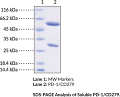
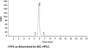
Technical Information
- CD279
- PDCD1
- Programmed Cell Death Protein 1
Shipping & Storage Information
Recommended Products
Certificates of Analysis & Batch Specific Data
Provide batch numbers separated by commas to download or request available product inserts, QC sheets, certificates of analysis, data packs, and GC-MS data.

Visit Our Cancer Resource Center
Find Tools & Resources to Study the Hallmarks of Cancer
- Cancer cell signaling & regulation
- Cancer metabolism
- Tumor microenvironment
Product Description
Programmed cell death protein 1 (PD-1), also known as CD279, is a cell surface receptor belonging to the immunoglobulin superfamily that is involved in regulation and attenuation of the adaptive immune response.1,2 It is a 288-amino acid type I transmembrane protein encoded by the PDCD1 gene in humans and is composed of a 167-amino acid ectodomain consisting of an N-loop, IgV-like domain, and a stalk region, a transmembrane domain, and a cytoplasmic tail with two tyrosine-based signaling motifs.3,4,2,1 PD-1 is expressed in activated immune cells, including CD4+ T cells, CD8+ T cells, natural killer T (NKT) cells, B cells, monocytes, and dendritic cells.5,2 Binding of PD-1 to either of its ligands, PD-L1 (Item No. 28378) or PD-L2 (Item No. 28379), suppresses T cell proliferation and cytokine production.1 PD-1 deficiency induces cardiomyopathy or lupus-like glomerulonephritis in BALB/c and C57Bl/6 mice, respectively.3,2 Increased levels of intratumoral PD-1+ immune cells are associated with increased tumor size, higher nuclear grade, and poor prognosis in patients with renal cell carcinoma.6 Formulations containing PD-1 blocking antibodies have been used in the treatment of various cancers. Cayman’s PD-1/CD279 Recombinant Monoclonal Antibody (Pembrolizumab) recognizes PD-1 from human samples.
WARNING This product is not for human or veterinary use.
References & Product Citations
1. PD-
2. PD-
3. PDCD1: A tissue-
4. An unexpected N-
5. PD-
6. PD-
Contact Info
Cayman Chemical1180 East Ellsworth RoadAnn Arbor, Michigan 48108 USAToll Free: (800) 364-9897(USA and Canada only)Fax: (734) 971-3640


