
News & Announcements
Sphingolipid Solubilization, Isolation, Analysis, and Storage Advice
Article from 2021-03-19
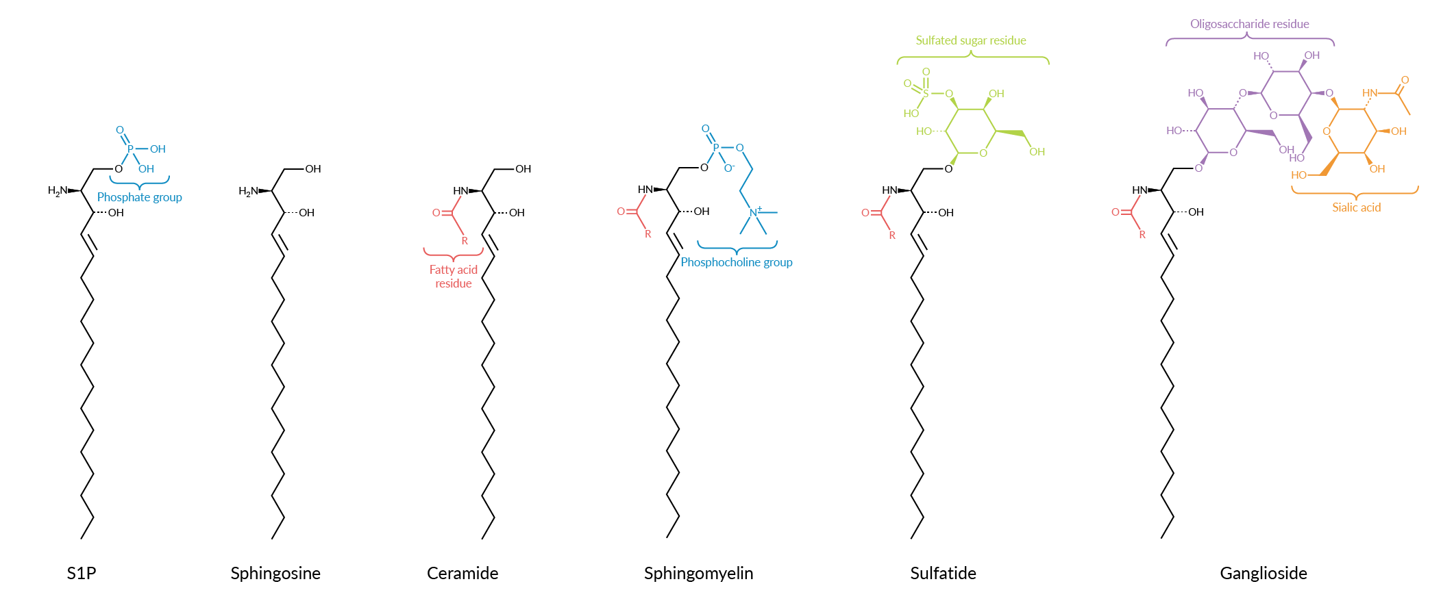
The information and methods below are designed to help you achieve the best results possible with our sphingolipid products. As much as we would like to guarantee individual success of the methods below, experimental nuances can sometimes interfere with the outcome. We want to help you advance your research goals. Our scientists are available to provide technical support, research tools, and services to help make your research possible. Feel free to contact them if you need further assistance in your lipid research. Contact a technical support scientist
Sphingolipid Solubility
Some lipids, such as sphingolipids, have limited or very poor solubility in many common solvents such as chloroform, hexane, ethyl ether, and even methanol. To overcome this insolubility problem, various solvent mixtures have been developed. One of the most universal sphingolipid solvents is a mixture of chloroform/methanol/water. However, while this solvent system is very useful for analytical testing, it is very toxic towards cells and cannot be used in live cell cultures or other in vivo applications. Several alternative methods have been developed to provide sphingolipid solutions suitable for live-cell studies. Although these methods were primarily developed for ceramides and glucosylceramides, they may be adapted for use with other lipids. Applicable solubility systems include:
Biological Applications:
Method A: BSA-Lipid Complexes
Prepare ~1 mM lipid stock solution in chloroform/methanol (19:1, v/v). Dispense 50 μl of the lipid stock solution into a glass test tube and dry, first under a stream of nitrogen, and then in vacuo for at least 1 hour. Redissolve the dried lipid in 200 μl ethanol. Mix 10 ml of buffer (100 mM NaH2PO4/Na2HPO4, pH 7.4) with 3.4 mg (0.34 mg/ml) of fatty acid-free BSA into a 50 ml plastic centrifuge tube.1,2 Agitate on a vortex mixer. Inject the 200 μl lipid solution into the BSA solution while vortexing. Store the resulting solution (5 μM lipid + 5 μM BSA) in a plastic tube at -20°C.
Method B: Solubilization Using Zwitterionic Detergent CHAPS
Evaporate a solution containing 15 nmol of lipid under a stream of nitrogen in a 1.5 ml test tube. Add a solution of 1.1 mg of CHAPS (3-[(3-cholamidopropyl)dimethylammonio]-1-propanesulfonate) in 10 μl of phosphate buffer (100 mM NaH2PO4/Na2HPO4, pH 7.4). Thoroughly mix and sonicate for 3 minutes.2
Method C: Solubilization Using Ethanol/Dodecane (98:2, v/v) for Use with Tissue Homogenates
Evaporate a solution containing 15 nmol of lipid under a stream of nitrogen in a 1.5 ml test tube. After dissolving in ethanol/dodecane (98:2, v/v; 1% final concentration), add phosphate buffer (100 mM NaH2PO4/Na2HPO4, pH 7.4) and sonicate the solution for 3 minutes.2
Method D: Solubilization Using Ethanol:Dodecane (98:2, v/v) for Use in Cell Cultures
Dissolve ceramide (from bovine brain sphingomyelin or cerebroside), C2-ceramide, DAG, or sphingosine in ethanol:dodecane (98:2 v/v). Add this solution to DMEM and Ham's F12 medium in a tube and mix well with the medium using agitation.3 Transfer to culture dishes.
Mass Spectrometry/Organic Chemistry Applications:
Method E: Hexane/Propanol
Dissolve the lipid in hexane/2-propanol (3:2, v/v). Can add 5.5% water. Can also add 0.01% BHT to limit oxidation.4 This method is best reserved for gas chromatography applications.
Universal Sphingolipid Solvent (this system will work for most sphingolipids)
Dissolve in chloroform/methanol (2:1, v/v) at a concentration of 10 mg lipid/ml solvent. If the lipid is still not soluble, add water up to 10% of the solvent volume. Sonication or mild heating (up to 40°C) can help to dissolve the lipid faster. A revised version of this solvent using 1:1 butanol/methanol may also be used.
Lipid | Applicable Solubility Systems* |
| Sphingosine Dihydrosphingosine Phytosphingosine Ceramide Dihydroceramide Phytoceramide |
|
| Sphingomyelin Sphingosylphosphorylcholine |
|
| Sphingosine-1-phosphate Ceramide-1-phosphate |
|
Glycosphingolipids: Galactosylceramides |
|
| Gangliosides |
|
*Please visit individual product pages to see specific solvents that we have tested with that particular product.
References
1. Pagano, R.E. A fluorescent derivative of ceramide: Physical properties and use in studying the Golgi apparatus of animal cells. Methods Cell Biol. 29, 75-85 (1989).
2. Michel, C., van Echten-Deckert, G., Rother, J., et al. Characterization of ceramide synthesis. A dihydroceramide desaturase introduces the 4,5-trans-double bond of sphingosine at the level of dihydroceramide. J. Biol. Chem. 272(36), 22432-22437 (1997).
3. Ji, L., Zhang, G., Uematsu, S., et al. Induction of apoptotic DNA fragmentation and cell death by natural ceramide. FEBS Lett. 358(2), 211-214 (1995).
4. Norm Radin (unpublished observations)
Sphingolipid Isolation
Due to the wide range of physical properties between various lipid species, it is difficult to isolate all lipids with one technique. Different processes are generally used to extract polar and non-polar compounds. Some methods allow for the isolation of all lipid species but are not necessarily as quantitative as more specialized protocols. Many references are available to choose from depending on your requirements. Two very helpful online resources to get familiar with various protocols are available at the AOCS Lipid Library and Cyberlipid Center.
The most common extraction techniques for lipids are the Folch and the Bligh and Dyer methods, which are outlined below:
Folch Method
Homogenize tissue sample with chloroform/methanol 2:1, 20 ml/gram of tissue.
Remove the solids by filtration or centrifugation.
Add 0.2 volumes of 0.9% KCl or NaCl.
Gently mix and allow the layers to separate. Centrifugation will speed up the separation.
Gangliosides are mostly present in the aqueous phase and can be recovered by reversed phase chromatography.
Almost all other lipids are in the organic solvent layer and can be concentrated and purified by normal phase chromatography.
Bligh and Dyer Method
Homogenize tissue sample with 1 ml of water, 1M NaCl, or 1 M KCl.
Add 3.75 ml chloroform/methanol 1:2 and homogenize.
Add 1.25 ml chloroform and mix.
Add 1.25 ml water and mix.
Centrifuge to separate the aqueous and organic phases.
Gangliosides are mostly present in the aqueous phase and can be recovered by reversed phase chromatography.
Almost all other lipids are in the organic solvent layer and can be concentrated and purified by normal phase chromatography.
Suggested References for Analytical Sphingolipid Extraction and Analysis
Glucosylsphingosine
Fuller, M., Szer, J., Stark, S., et al. Rapid, single-phase extraction of glucosylsphingosine from plasma: A universal screening and monitoring tool. Clin. Chim. Acta 450, 6-10 (2015).
Sulfatides
Barcenas, M., Suhr, T.R., Scott, C.R., et al. Quantification of sulfatides in dried blood and urine spots from metachromatic leukodystrophy patients by liquid chromatography/electrospray tandem mass spectrometry. Clin. Chim. Acta 433, 39-43 (2014).
Gangliosides
Masson, E.A.Y., Sibille, E., Martine, L., et al. Apprehending ganglioside diversity: A comprehensive methodological approach. J. Lipid Res. 56(9), 1821-1835 (2015).
Zhang, Y., Wang, J., Liu, J. et al. Combination of ESI and MALDI mass spectrometry for qualitative, semi-quantitative and in situ analysis of gangliosides in brain. Sci. Rep. 6, 25289 (2016).
Tropak, M.B., Yonekawa, S., Karumuthil-Melethil, S., et al. Construction of a hybrid β-hexosaminidase subunit capable of forming stable homodimers that hydrolyze GM2 ganglioside in vivo. Mol. Ther. Methods Clin. Dev. 3, 15057 (2016).
Fatty Acid Analysis of Glycerides, Phospholipids, and Sphingolipids by GC/FID
Natural sphingolipids contain a heterogeneous mixture of fatty acids attached to the sphingosine backbone. The fatty acid composition is dependent on several factors including species, location within the organism, age, and environmental conditions. Normal variations in the fatty acids include chain-length, hydroxylation, and unsaturation. To determine the fatty acid components present in a sphingolipid sample, the fatty acid is hydrolyzed from the sphingosine backbone and then converted to a fatty acid methyl ester. It can then be analyzed by GC using flame ionization detection (FID), and the individual fatty acid methyl esters identified by comparison of the retention times to reference standards.
Preparation of Methyl Esters from Glycerides and Phospholipids
Methanolic Base Hydrolysis
Weigh 5 mg of dry sample to be tested into a reaction vessel.
Add 1 ml of hexane to the sample.
Add 50 μl of 1 M sodium methoxide in methanol.
Mix thoroughly.
Let sit at room temperature for 5 minutes.
The sample is now ready to analyze by GC/FID.
Preparation of Methyl Esters from Phospholipids, Neutral Sphingolipids, Neutral Glycosphingolipids, and Acidic Glycosphingolipids
Methanolic Sulfuric Acid
Make a solution of 2% sulfuric acid in methanol. The 2% sulfuric acid/methanol solution must be made fresh before use.
Add 1 ml of the sulfuric acid solution to the dry lipid sample in a reaction tube.
Blanket with argon and seal with a Teflon-lined cap.
Place in an equilibrated heating block as indicated below:
Fatty acid to methyl ester – 5 minutes at 80°C
2- or 3-Hydroxy fatty acids – 1.5 hours at 80°C
Phospholipids – 2 hours at 70°C
Glycolipids – 6 hours at 100°C
Sphingomyelin – 2 hours at 100°C
At the end of the desired time, remove from the heating block and let cool to room temperature. Do not open until cool!
Add 0.5 ml of deionized water, which will stop the reaction.
Add 4 ml of hexane and shake vigorously.
Let stand until two phases are visible. Note: Top hexane layer must be clear.
Remove the top hexane layer and save.
Extract the bottom layer by repeating steps 7-9 two more times.
Discard the bottom layer after fully extracted.
Combine the three top layers and add a 0.5-1 g of a mixture of sodium sulfate/ sodium bicarbonate (4:1).
Shake vigorously.
Using a pipette, transfer the sample to a reaction tube. Note: Be careful not to pull any of the sodium sulfate/sodium bicarbonate solids.
Concentrate with a stream of nitrogen down to 1 ml.
The sample is now ready to analyze by GC/FID.
GC/FID Analysis of Fatty Acid Methyl Esters
GC Conditions*
Column: SP-2330 or RTX-2330; 30 x 0.25 mm x 0.2 μm
Oven: 200°C Isothermal
Carrier Linear Velocity: Helium at 20 cm/sec.
Detector: FID, 250°C
Injector: 250°C
Split ratio: 100:1
*These are general GC conditions for fatty acid methyl esters and may not separate all components of a sample.
Storage and Handling of Lipids
Natural lipid products can contain significant amounts of unsaturation, both in the fatty acid moieties and elsewhere. These are prone to autoxidation when exposed to oxygen, water, or other oxidants. Temperature can also significantly impact the rate of oxidation. Unsaturated lipids should be handled with extra precautions.
Catalog items in unopened containers are generally stable for at least one year when stored under the conditions indicated in the catalog listing. Items containing unsaturated fatty acids are especially subject to oxidation and should be stored in a solution of organic solvents, or under argon, at -20°C. Minimize exposure to air for these products. Glycolipids and phospholipids should not be stored in aqueous solutions due to potential hydrolysis.
Never use plastic or polymer materials with organic solvents as doing this will leach out impurities from the material. Always use glass, stainless steel, or Teflon equipment for transferring lipids in organic solvents and for storage. Lipids should always be stored in glass containers with Teflon lined caps.
You May Also Be Interested In
| Eicosanoid Sample Collection, Preparation, and Storage Advice | Meet the Expert: Lipidomic Profiling Services |
 |  |
| Analytical Techniques | Sphingolipid Shorthand |
 |  |
Contact Info
Cayman Chemical1180 East Ellsworth RoadAnn Arbor, Michigan 48108 USAToll Free: (800) 364-9897(USA and Canada only)Fax: (734) 971-3640


