
News & Announcements
Glucosylsphingosine: A Sensitive and Specific Biomarker for Gaucher Disease
Article from 2018-08-01
This article was originally published in the August 2018 edition of Matreya’s Newsletter for Glyco/Sphingolipid Research (PDF).
Lysosomal storage diseases are a heterogeneous group of disorders caused by lysosomal enzyme dysfunction. Gaucher disease (GD) is the most common of these lysosomal storage disorders and has recently warranted much research due to the debilitating effects of excess lipid storage in Gaucher cells. A lack of activity in the lysosomal enzyme glucosylceramidase (GCase), or occasionally in its activator protein saposin C, causes an accumulation of glucosylceramide, glucosylsphingosine, and other glycosphingolipids in macrophage cells, especially in the liver, spleen, lung, and bone marrow. These lipid-heavy cells are commonly known as "Gaucher cells" and can result in hepatosplenomegaly, cytopenia, skeletal disfunctions, lung disorders, and neuronal degradation.
Chitotriosidase is the most well-established biomarker for GD. However, it is not specific for GD and may give a false negative in a significant percentage of patients due to a particular mutation. The incidence of deficiency of chitotriosidase activity recorded in the Netherlands and among the Ashkenazi Jewish population predicts that 37% of the population will be carriers of a mutant chitotriosidase gene.1 Furthermore, chitotriosidase also reflects the changes in the course of the disease belatedly. Due to these limitations, a more specific biomarker is needed for GD.
A report by Elstein et al. demonstrated that glucosylsphingosine can be used as a reliable and specific biomarker for GD as well as for measuring the effectiveness of enzyme replacement therapy (ERT).2 Evaluation of the sensitivity and specificity of glucosylsphingosine as a biomarker during ERT showed significantly decreased levels of glucosylsphingosine in both new patients and patients that had previously been treated using ERT. The quantitation of glucosylsphingosine was demonstrated to serve as a direct indicator of disease intensity and response to ERT.2
Taking advantage of glucosylsphingosine as a biomarker, Fuller et al. developed a quick and reproducible method for the determination of abnormally high glucosylsphingosine levels from 0.01 ml of plasma.3 With this method, plasma is spiked with N-palmitoyl-lactosylceramide-d3 as an internal standard and extracted with chloroform/methanol. LC/ESI-MS/MS analysis showed recovery to be >90%, and the calibration curve was linear over the entire relevant range. The assay was described as "accurate, reproducible, robust, and easy to perform in routine laboratory settings." This method found that glucosylsphingosine was elevated in all GD patients compared to unaffected controls and patients with other metabolic disorders.
Cayman offers 13C6 glucosylsphingosine (d18:1) and N-Glycine glucosylsphingosine (d18:1) as highly specific glucosylsphingosine internal standards. 13C6 Glucosylsphingosine (d18:1) contains six carbon-13 units on the glucose moiety while N-glycine glucosylsphingosine (d18:1) contains a glycine attached to the amine of sphingosine, preserving its primary amine. These sphingolipids are ideal for use as internal standards in the extraction and mass spectrometry analysis of samples. The free amine group of glycine gives very similar physical characteristics to the natural sphingolipid, while the glycine adds an additional 57 Da to the molecule, making it easy to detect by mass spectroscopy methods.4
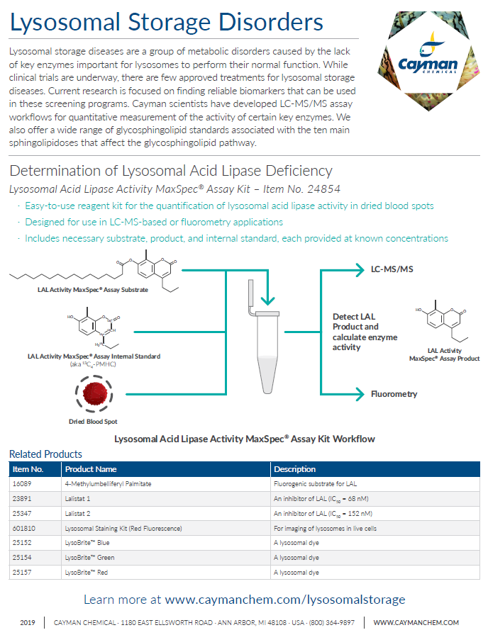
More Lysosomal Storage Disorders Research Tools from Cayman
Further Reading
Lysosphingomyelin: A Sensitive and Specific Biomarker for Niemann-Pick Disease
References
1. Boot, R.G., Renkema, G.H., Verhoek, M., et al. The human chitotriosidase gene. Nature of inherited enzyme deficiency. J. Biol. Chem. 273(40), 25680-25685 (1998).
2. Elstein, D., Mellgard, B., Dinh, Q., et al. Reductions in glucosylsphingosine (lyso-Gb1) in treatment-naïve and previously treated patients receiving velaglucerase alfa for type 1 Gaucher disease: Data from phase 3 clinical trials. Mol. Genet. Metab. 122(1-2), 113-120 (2017).
3. Fuller, M., Szer, J., Stark, S., et al. Rapid, single-phase extraction of glucosylsphingosine from plasma: A universal screening and monitoring tool. Clin. Chim. Acta 450, 6-10 (2015).
4. Krüger, R., Tholey, A., Jakoby, T., et al. Quantification of the Fabry marker lysoGb3 in human plasma by tandem mass spectrometry. J. Chromatogr. B Analyt. Technol. Biomed. Life Sci. 883-884, 128-135 (2012).
Contact Info
Cayman Chemical1180 East Ellsworth RoadAnn Arbor, Michigan 48108 USAToll Free: (800) 364-9897(USA and Canada only)Fax: (734) 971-3640


