
News & Announcements
Advancing MASLD & MASH Research: PPAR and FFAR Fatty Acid Signaling Pathways
Article from 2024-08-28
Previous Post: Advancing MASLD & MASH Research: Targeting De Novo Lipogenesis & Insulin Resistance
Metabolic dysfunction-associated steatotic liver disease (MASLD) and metabolic dysfunction-associated steatohepatitis (MASH) are complex steatotic liver diseases (SLDs). At the heart of these conditions are metabolic disturbances, with lipid homeostasis playing a crucial role. One way to maintain lipid homeostasis is by targeting peroxisome proliferator-activated receptors (PPARs) and free fatty acid receptors (FFARs) in MASLD and MASH.
Regulating Lipid Homeostasis by Targeting Fatty Acid Signaling Pathways
PPARs
Peroxisome proliferator-activated receptors (PPARs) are ligand-regulated transcription factors that act as central regulators of glucose, lipid, and cholesterol homeostasis. Each PPAR subtype exhibits distinct roles and cell/tissue expression levels that complement eachother.1-3 These nuclear receptors also crosstalk with inflammatory pathways, offering a double-pronged therapeutic approach that not only addresses metabolic dysfunction but also inflammation in MASLD and MASH.4
Effects of PPAR Agonists
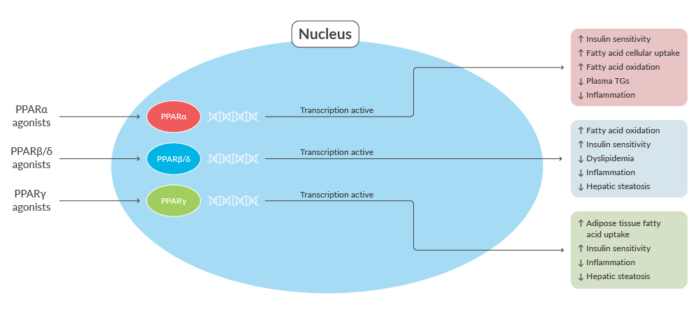
Strategies to Target PPARs | ||
| Class | Effects | References |
| PPARα agonists PPARα is highly expressed in cells and tissues that oxidize fatty acids at a rapid rate (e.g., liver, brown adipose tissue, heart, kidney, and skeletal muscle) and has a major role in fatty acid metabolism. | ↑ Insulin sensitivity | 1,2,4-7 |
| PPARβ/δ agonists PPARβ/δ is expressed extensively, with high expression levels in the liver, adipose tissue, and skeletal muscle. It has major roles in fatty acid metabolism, glucose homeostasis, and the regulation of cholesterol levels. | ↑ Fatty acid oxidation ↑ Insulin sensitivity ↓ Dyslipidemia ↓ Inflammation ↓ Hepatic steatosis | 1,2,4-7 |
| PPARγ agonists PPARγ is broadly expressed, with high expression levels in liver, white adipose tissue, macrophages, and skeletal muscle. It has central roles in lipogenesis and glucose homeostasis. | ↑ Adipose tissue fatty acid uptake | 2,4-8 |
Assay Kits for PPAR Signaling: Transcription Factor Assay Kits & Cell-based Reporter Assays
Transcription factor assay kits measure the relative abundance of the transcription factor in a sample whereas cell-based reporter assays quantify the transcriptional output of a transcription factor with a reporter gene.
PPAR Transcription Factor Assay Kits
| Item No. | Product Name | Description |
| 10006855 | PPARγ Transcription Factor Assay Kit | A 96-well assay for measurement of PPARγ DNA binding activity |
| 10006914 | PPARδ Transcription Factor Assay Kit | A 96-well assay for measurement of PPARδ DNA binding activity |
| 10006915 | PPARα Transcription Factor Assay Kit | A 96-well assay for measurement of PPARα DNA binding activity |
| 10008878 | PPARα, δ, γ Complete Transcription Factor Assay Kit | A 96-well assay for measurement of PPARα, δ, and γ |
PPAR Cell-based Reporter Assays
| Item No. | Product Name | Description |
| 15732 | Human Peroxisome Proliferator-Activated Receptor Panel | A nuclear receptor cell-based reporter assay |
| 15729 | Human Peroxisome Proliferator-Activated Receptor Gamma Reporter Assay System | A nuclear receptor cell-based reporter assay |
| 15730 | Human Peroxisome Proliferator-Activated Receptor Alpha Reporter Assay System | A nuclear receptor cell-based reporter assay |
| 15731 | Human Peroxisome Proliferator-Activated Receptor Beta/Delta Reporter Assay System | A nuclear receptor cell-based reporter assay |
View all PPAR cell-based reporter assays
Worth noting, there are also species differences in PPAR physiology between rodents and humans, and there are limitations with applying preclinical data obtained from rodent models to humans.5
FFARs
Like PPARs, FFA receptors (FFARs) bind fatty acids and regulate signaling pathways related to hormone secretion, carbohydrate and lipid metabolism, and immune responses.9,10 However, while PPARs are ligand-activated transcription factors that regulate gene transcription, FFARs are cell surface receptors that belong to the G protein-coupled receptor (GPCR) family and initiate intracellular signaling cascades.9,10
There are several FFAR subtypes, including FFAR1 (GPR40), FFAR2 (GPR43), FFAR3 (GPR41), and FFAR4 (GPR120), which are expressed in different tissues and have distinct functions.9,10 In general, FFAR1 and FFAR4 preferentially bind medium-chain fatty acids (MCFAs) and long-chain fatty acids (LCFAs), whereas FFAR2 and FFAR3 preferentially bind short-chain fatty acids (SCFAs) produced via fermentation of dietary fiber by gut microbiota.9,10
Effects of FFAR Agonists
Strategies to Target FFARs | ||
| Class | Effects | References |
| FFAR1 agonists FFAR1 is expressed by enteroendocrine cells, pancreatic islet cells, immune cells, and hepatocytes.10 It senses FFAs to increase glucose-stimulated insulin secretion and the release of incretin hormones. | ↑ Glucose-stimulated insulin secretion ↑ GLP-1 and GIP secretion ↑ M2 macrophage polarization | 9-11 |
| FFAR2 agonists FFAR2 is expressed by adipocytes, enteroendocrine cells, pancreatic islet cells, and immune cells. Notably, it is not expressed in the liver. FFAR2 regulates lipid metabolism and glucose homeostasis. | ↑ GLP-1 secretion ↑ Insulin secretion ↓ Lipolysis ↓ Lipid accumulation ↓ Inflammation | 9,10 |
| FFAR3 agonists FFAR3 is expressed by adipocytes, enteroendocrine cells, and pancreatic islet cells. Notably, FFAR3 is not expressed in the liver. FFAR3 has important roles in lipid metabolism and glucose homeostasis. | ↑ GLP-1 secretion ↑ Energy expenditure | 9,10 |
| FFAR4 agonists FFFAR4 is expressed by adipocytes, enteroendocrine cells, immune cells, and hepatocytes. FFAR4 regulates hormone secretion in the intestine and pancreas. | ↑ Insulin secretion ↑ GLP-1 and GIP secretion ↓ Inflammation ↑ Insulin sensitivity ↓ Hepatic steatosis | 9,10 |
Assay Kits for FFARs
| Item No. | Product Name | Description |
| 601190 | FFAR1 (GPR40) Reporter Assay Kit | A reverse transfection reporter assay to screen for FFAR1 agonists, antagonists, and modulators |
| 601200 | FFAR4 (GPR120) Reporter Assay Kit | A reverse transfection reporter assay to screen for FFAR4 agonists, antagonists, and modulators |
The many roles of these receptors in fatty acid signaling pathways related to lipid homeostasis, glucose metabolism, and inflammation make them attractive targets for therapeutic approaches. These approaches not only address lipid accumulation but also have the potential to improve insulin sensitivity and reduce inflammation, addressing multiple facets of MASLD pathogenesis.
Lipidomics & Lipid Analysis Services
Cayman also offers a suite of services that may be of interest to MASLD and MASH researchers. Cayman's Lipidomics & Lipid Analysis Services offer researchers the opportunity to analyze lipid profiles in biological samples with our state-of-the-art facilities and of decades of collective expertise in lipid synthesis, purification, and characterization.
However, another aspect of lipid homeostasis with important implications in MASLD and MASH is cholesterol metabolism and how that relates to signaling pathways mediated by liver X receptors (LXRs), a subset of nuclear receptors that play a pivotal role in cholesterol homeostasis and lipid metabolism, and receptors for bile acids, potent signaling molecules that influence cholesterol homeostasis.
In the next section, explore how targeting cholesterol metabolism through modifications in LXR and bile acid receptor signaling could offer new approaches for MASLD and MASH.
Next Post: Advancing MASLD & MASH Research: The Roles of LXRs, FXRs, and GP-BAR1 in Metabolic Regulation
Related Posts:
- Advancing MASLD & MASH Research: An Introduction
- Advancing MASLD & MASH Research: Targeting De Novo Lipogenesis & Insulin Resistance
Products for MASLD & MASH Research
The products featured in this series are a snapshot of the comprehensive resources available from Cayman for MASLD and MASH research. Our full catalog contains a comprehensive range of biochemicals, proteins, antibodies, and assay kits to support MASLD and MASH research.
View all MASLD-related products
References
1. Puengel, T., Liu, H., Guillot, A., et al. Nuclear receptors linking metabolism, inflammation, and fibrosis in nonalcoholic fatty liver disease. Int. J. Mol. Sci. 23(5), 2668 (2022).
2. Yang, Z., Danzeng, A., Liu, Q., et al. The role of nuclear receptors in the pathogenesis and treatment of non-alcoholic fatty liver disease. Int. J. Biol. Sci. 20(1), 113-126 (2024).
3. Christofides, A., Konstantinidou, E., Jani, C., et al. The role of peroxisome proliferator-activated receptors (PPAR) in immune responses. Metabolism 114, 154338 (2021).
4. Tyagi, S., Gupta, P., Saini, A.S., et al. The peroxisome proliferator-activated receptor: A family of nuclear receptors role in various diseases. J. Adv. Pharm. Technol. Res. 2(4), 236-240 (2011).
5. Liss, K.H.H. and Finck, B.N. PPARs and nonalcoholic fatty liver disease. Biochimie 136, 65-74 (2017).
6. Grygiel-Górniak, B. Peroxisome proliferator-activated receptors and their ligands: Nutritional and clinical implications - a review. Nutr. J. 13, 17 (2014).
7. Lange, N.F., Graf, V., Caussy, C., et al. PPAR-targeted therapies in the treatment of non-alcoholic fatty liver disease in diabetic patients. Int. J. Mol. Sci. 23(8), 4305 (2022).
8. Chandra, M., Miriyala, S., and Panchatcharam, M. PPARγ and its role in cardiovascular diseases. PPAR Res. 6404638 (2017).
9. Grundmann, M., Bender, E., Schamberger, J., et al. Pharmacology of free fatty acid receptors and their allosteric modulators. Int. J. Mol. Sci. 22(4), 1763 (2021).
10. Secor, J.D., Fligor, S.C., Tsikis, S.T., et al. Free fatty acid receptors as mediators and therapeutic targets in liver disease. Front. Physiol. 12, 656441 (2021).
11. Kumari, P., Inoue, A., Chapman, K., et al. Molecular mechanism of fatty acid activation of FFAR1. Proc. Natl. Acad. Sci. USA 120(22), e2219569120 (2023).
Contact Info
Cayman Chemical1180 East Ellsworth RoadAnn Arbor, Michigan 48108 USAToll Free: (800) 364-9897(USA and Canada only)Fax: (734) 971-3640


