
News & Announcements
New Product Spotlight: Biofilm Formation and Staining Kit
Article from 2025-11-11
Cayman Chemical's new Biofilm Formation and Staining Kit (Item No. 502869) provides a straightforward, semi-quantitative method for evaluating bacterial biofilm formation. This assay kit allows you to easily screen for mutations and/or compounds that modulate biofilm formation with less time and variability than traditional staining methods.
About Bacterial Biofilms
Many species of bacteria possess the ability to adhere to biological or synthetic surfaces and form multicellular communities known as biofilms. An estimated 65-80% of bacterial infections are associated with biofilms, including chronic wound infections, endocarditis, otitis media, medical implant contamination, and chronic lung infections in patients with cystic fibrosis.1,2 Major human pathogens known to cause biofilm-associated disease include Proteus mirabilis, which can cause catheter-associated urinary tract infections, as well as Escherichia coli, Pseudomonas aeruginosa, and Staphylococcus aureus.1-4
Within a biofilm, bacteria are encased in an extracellular matrix (ECM) comprised of exopolysaccharides (EPS), proteins, lipids, and nucleic acids that protects them from clearance by the host immune system or antibiotics (Figure 1).1,2 In fact, bacteria are 1,000 times more tolerant to antibiotics when they are encased in a biofilm compared with existing in a motile, planktonic state.1
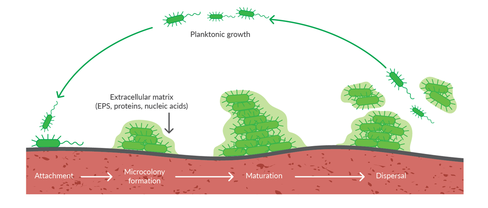
Figure 1. Bacterial biofilm formation and dispersal. Following attachment of motile, planktonic bacteria to a surface, cell division and production of extracellular matrix components leads to formation of microcolonies that mature into complex biofilms. Individual cells can disperse from the biofilm to resume a planktonic lifestyle or sections of the biofilm can be dispersed by mechanical disruption.
Finding new ways to inhibit biofilm formation or persistence may help facilitate prevention or treatment of biofilm-associated infections. Cayman's new Biofilm Formation and Staining Kit provides a straightforward, reliable method to screen for mutations and/or compounds that modulate bacterial biofilm formation.
About the Assay
Cayman's Biofilm Formation and Staining Kit provides a colorimetric semi-quantitative method for evaluating biofilm production on polystyrene pegs (Figure 2). Growth of biofilm on peg lids significantly reduces wash time and decreases variability. In the assay, crystal violet binds to negatively charged molecules within the ECM secreted by bacteria during biofilm formation. The absorbance of crystal violet can be measured at 590 nm. The amount of bound dye is proportional to the biofilm mass. This kit provides reagents sufficient to assay 20 samples in triplicate.
Figure 2. Assay scheme
See the Assay in Action
| |
Kit Performance
Decreased Variability
Cayman's assay kit reduces variability compared to traditional assay methods. When used to examine biofilm formation by P. mirabilis, Cayman's peg lid-based assay kit demonstrated a coefficient of variance of only 6.3%, compared to 16.4% obtained using a traditional method (Figure 3).
Figure 3. Assay performance of peg-based assay compared to traditional assay methodology. Culture plates were inoculated with 1 x 10⁷ CFU/ml of P. mirabilis strain HI4320 and incubated statically for 24 hours at 37°C with either a 96-peg lid or a traditional 96-well lid prior to staining.
Species Versatility
Biofilm composition varies by bacterial species. When tested using Gram-positive cocci (S. saprophyticus) and Gram-negative bacilli (E. coli and P. mirabilis) bacteria, this assay successfully stained all three species (Figure 4).
Figure 4. Versatility of the Biofilm Formation and Staining Kit. Bacteria were inoculated at 1 x 10⁷ CFU/ml and incubated statically for 24 hours prior to staining.
Detection of Biofilm-Defective Mutants
We also assessed whether the Biofilm Formation and Staining Kit could identify genes of interest in biofilm formation and/or regulation. To examine this, we used the assay kit to measure biofilm formation in wild-type P. mirabilis HI4320 and strains possessing mutations in genes implicated in biofilm formation (crp, bcsA, bcsA2, and bcsB2).5,6
CRP together with its cofactor cyclic AMP (cAMP-CRP) can promote or inhibit biofilm formation, depending on the species of bacteria in question.5 BcsA and BcsB, subunits of the bacterial cellulose synthase complex, are essential for the production of cellulose, which is a significant component of the ECM in bacterial biofilms.6
Using the Biofilm Formation and Staining Kit, we demonstrated that P. mirabilis HI4320 possessing transposon insertion mutations in crp, bcsA, bcsA2, and bcsB2 showed statistically significant decreases in biofilm formation compared to the wild-type (WT) strain (Figure 5).
Figure 5. Defective biofilm formation in P. mirabilis HI4320 mutants. Wild-type (WT) P. mirabilis HI4320 and various transposon insertion mutants were grown statically in LB for 24 hours at 37°C to allow biofilm formation. Biofilm biomass was determined using the Biofilm Formation and Staining Kit. Absorbance at 590 nm was normalized to total bacterial growth (OD600) and then to WT. Statistical significance was determined by one-way ANOVA; p<0.05 (*), p<0.001 (**).
For additional information on the performance of this kit, please refer to the kit booklet (PDF).
The Cayman Advantage
At Cayman, we have an exceptional understanding of assay development, validation, and performance. Each of our assay kits undergoes rigorous quality testing to certify high precision and accuracy. Our attention to these details ensures you will obtain reproducible results, from day to day and lot to lot, with expert technical support readily available to assist you. Learn more about the Cayman Advantage in our article, Why Cayman Assay Kits.
Our Biofilm Formation and Staining Kit is also complemented by our line of cyclic dinucleotide (CDN) ELISA kits that enable sensitive, specific detection of these cellular second messengers, several of which regulate biofilm formation in bacteria. Explore the resources below to discover additional resources to study bacterial physiology, biofilm formation, and infectious disease.
You May Also Be Interested In
| |
|
Quantification of Cyclic Dinucleotides During Growth Phase in Three Bacterial Species Using ELISAs |
References
1. Sambanthamoorthy, K., Sloup, R.E., Parashar, V., et al. Identification of small molecules that antagonize diguanylate cyclase enzymes to inhibit biofilm formation. Antimicrob. Agents Chemother. 56(10), 5202-5211 (2012).
2. Martín-Rodríguez, A.J. and Römling, U. Nucleotide second messenger signaling as a target for the control of bacterial biofilm formation. Curr. Top. Med. Chem. 17, 1928-1944 (2017).
3. Yan, J. and Bassler, B.L. Surviving as a community: Antibiotic tolerance and persistence in bacterial biofilms. Cell Host Microbe 26(1), 15-21 (2019).
4. Kynshi, M.A.L., Kharkamni, E., and Borah, V.V. Proteus mirabilis: Insights into biofilm formation, virulence mechanisms, and novel therapeutic strategies. Microbe 8, 100450 (2025).
5. Liu, C., Sun, D., Zhu, J., et al. The regulation of bacterial biofilm formation by cAMP-CRP: A mini-review. Front. Microbiol. 11, 802 (2020).
6. Römling, U. and Galperin, M.Y. Bacterial cellulose biosynthesis: Diversity of operons, subunits, products, and functions. Trends Microbiol. 23(9), 545-557 (2015).
Contact Info
Cayman Chemical1180 East Ellsworth RoadAnn Arbor, Michigan 48108 USAToll Free: (800) 364-9897(USA and Canada only)Fax: (734) 971-3640


