
News & Announcements
Screening Inhibitors of Citrullination
Screening Inhibitors of Citrullination
Article from 2017-06-20
Citrullination—the process of deimination of charged arginine residues to neutral citrulline residues within proteins—has been linked to numerous inflammatory and autoimmune disorders, necessitating the search for biomarker assays and potent, targeted inhibitors of this post-translational modification for therapeutic benefit. Indeed, the first antibodies against citrullinated proteins were identified in rheumatoid arthritis (RA) patients in 1998.1 This ultimately led to a diagnostic test for anti-citrullinated protein antibodies (ACPA) in patients suspected to have RA. Since then, the presence of citrullinated peptides has also been linked to cancer, systemic lupus erythematosus, multiple sclerosis, and Alzheimer's disease.
While the introduction of citrulline in a protein—which can dramatically alter its structure and function—is implicated in pathological processes, it is important for several physiological functions as well, including epithelial terminal differentiation, gene expression regulation, apoptosis, and neutrophil-mediated host defense. For example, the citrullination of histones enables neutrophils to form web-like extruded complexes consisting of DNA and antimicrobial agents, neutrophil extracellular traps (NETs), to ensnare certain pathogens.2 That is, the citrullination of histones during NET formation helps reduce histone binding affinity for DNA and promote the decondensation of chromatin so that a web of double-stranded DNA (dsDNA) studded with histones, antimicrobial enzymes, and defensin peptides can be thrust out from neutrophils to capture, kill, and phagocytose bacteria and other pathogens.3-5 Once released, NET clearance is an equally important process since excessive circulating NET fragments could stimulate autoantibody production, as in the pathogenesis of systemic lupus erythematosus, which is characterized by the accumulation of autoantibodies to dsDNA and histones.6
The citrullination process is catalyzed by peptidylarginine deiminase (PAD) enzymes, whose activity is largely restricted to specific tissues and is regulated transcriptionally, translationally, and by the availability of calcium.7 Of the five known PAD isotypes, PAD2 and PAD4 are the most studied due to their impact in inflammation, histone modification, gene regulation, and autoimmune diseases. PAD2 is ubiquitously expressed in immune cells, skeletal muscle, spleen, brain, secretory glands, etc., and PAD4 is primarily restricted to neutrophils and eosinophils. Overexpression and increased activity of PAD2 and PAD4 have been described in several inflammatory and autoimmune diseases, as well as cancer.8-13
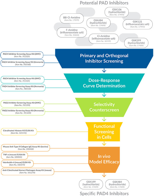
Because selective inhibition of PAD2 and PAD4 may prove to be a therapeutic target for certain human disorders, Cayman Chemical has developed a complete line of assays to enable the discovery of novel inhibitors of these enzymes. Antibodies, recombinant proteins, and a selection of inhibitors and probes have been developed to aid in evaluating the expression and activity of PAD in cells and in vivo efficacy models. Here we outline a logical screening workflow that exemplifies the use of Cayman’s assays to narrow down specific PAD4 inhibitors from a large library of potential compounds in the pre-clinical pursuit of effective disease treatments (Figure 1). Though the example is specific for PAD4, a parallel set of screening assays are also available to specifically evaluate PAD2 inhibitor activity.
(Click to Download PDF)
Figure 1. Pre-clinical screening funnel to identify selective PAD4 inhibitors.
Primary Inhibitor Screening
Cayman’s PAD4 Inhibitor Screening Assay Kit (AMC) provides a convenient method to initially identify small molecules that inhibit human PAD4. This assay utilizes a fluorescent substrate consisting of a modified arginine residue coupled to a 7-amino-4-methylcoumarin (AMC) fluorophore. Acylation of AMC onto the arginine residue masks the fluorescence of the AMC. In the absence of PAD4, the substrate remains unaltered, allowing the developer to release free AMC. In the presence of PAD4, the arginine of the substrate is citrullinated, and when the reaction is quenched by the addition of the developer, free AMC is prevented from release. In this reaction, the fluorescent signal is inversely proportional to the amount of citrullination by PAD4. Thus, a more intense signal indicates a greater degree of inhibition, allowing inhibitory compounds to be easily confirmed by visualizing a fluorescent signal.
Orthogonal Inhibitor Screening
Because false positives are inevitable in every high-throughput screening campaign, orthogonal assays are recommended to confirm hits and eliminate errors. Cayman’s PAD4 Inhibitor Screening Assay Kit (Ammonia) provides an alternative method for screening human PAD4 inhibitors. In this assay, ammonia is produced when PAD4 deiminates N-α-benzoyl-L-arginine ethyl ester, a non-natural substrate with similar kinetic properties to PAD’s natural substrates. Ammonia reacts with a detector, resulting in a fluorescent product. A negative result in this orthogonal assay would indicate that the primary hit was almost certainly an assay format-dependent artifact and not specific to the inhibition of PAD4. Compounds found to be active in both the AMC and ammonia formats are candidates for further analysis.
Specificity and Selectivity Determination
Either the PAD4 Inhibitor Screening Assay Kit (AMC) or the PAD4 Inhibitor Screening Assay Kit (Ammonia) can once again be used to perform dose-response curve (DRC) experiments. Compounds must be tested over a wide range of concentrations to determine the concentration that results in half-maximal activity (IC50). Subsequently, selectivity of the candidate small molecules for PAD4 over PAD2 can be tested with Cayman’s PAD2 Inhibitor Screening Assay Kit (AMC) or Cayman’s PAD2 Inhibitor Screening Assay Kit (Ammonia). This will allow for determination of compounds that show minimal potency towards an alternative target.
Cell-based, Functional Screening
Secondary screening of confirmed hits should next be tested in a functional cellular assay to determine efficacy. Activity in this secondary screen will confirm that compounds are able to function in a more complex biological system as opposed to the simple, isolated recombinant PAD4 protein used in the primary screen. HL-60 cells are one such model system that can be induced with DMSO to differentiate along the granulocyte lineage and express higher levels of PAD4. Upon calcium ionophore treatment, increased levels of PAD4-dependent histone citrullination are observed in these cells.14 Alternatively, histone citrullination can be observed in primary human neutrophils treated with a variety of stimuli.15 The measurement of citrullinated histones in this system can be a convenient cellular assay to test PAD4 inhibition.3,14,16-17 Cayman’s Citrullinated Histone H3 ELISA Kit measures citrullination at residues R2, R8, and R17 on histone H3 from cell lysates and, thus, can be used for the functional readout of pharmacological PAD inhibition in differentiated HL-60 cells or human primary neutrophils.
PAD-dependent Efficacy Models
Prior to lead optimization, work must be performed to examine compound efficacy in an appropriate in vivo model. Recently, a novel PAD4-selective inhibitor, GSK199 (hydrochloride), was shown to be effective in the mouse collagen-induced arthritis (CIA) model of RA.18 Cayman offers a series of products designed to support a PAD-dependent efficacy model of CIA. Cayman’s Mouse Anti-Type II Collagen IgG Assay Kit (bovine) is an immunometric assay that can be used to measure anti-type II collagen antibody in plasma or serum. This kit uses a bovine collagen-coated plate and an affinity-purified polyclonal antibody isolated from mice with CIA as a standard to provide a highly accurate measurement of anti-CII concentration in experimental plasma samples. A number of pro- and anti-inflammatory cytokines, including TNF-α and interleukin-6, are expressed in the joints of mice with CIA. These cytokines can be measured using Cayman’s TNF-α (mouse) ELISA Kit and Interleukin-6 (mouse) ELISA Kit.
As an alternative to the CIA model, mice (especially those expressing the human HLA-DR4 transgene) can be immunized with citrullinated human fibrinogen to produce an arthritic response driven by the production of antibodies that recognize citrullinated epitopes. The polyclonal antibody response produces antibodies reactive with both citrullinated human fibrinogen and unmodified, non-citrullinated human fibrinogen. Cayman’s Anti-Citrullinated Human Fibrinogen Assay Kit (mouse) is an immunometric assay that can be used to distinguish the antibody response to citrullinated human fibrinogen from the antibody response to unmodified human fibrinogen in mouse serum or plasma. A human fibrinogen affinity sorbent is provided with the kit so that any antibodies capable of reacting with non-citrullinated (unmodified) fibrinogen can be removed prior to analysis of the remaining anti-citrullinated fibrinogen antibodies for an accurate analysis of the anti-citrulline response.
Ready for Lead Optimization
By utilizing a systemic battery of tests as outlined above, select molecules can be swiftly sifted from a library of candidates for further optimization. Cayman has created this specific line of assays to give you the accuracy and efficiency needed to identify specific PAD inhibitors. We also offer full-service contract screening and profiling, including lead optimization and development, to help you identify particular modulators of PAD2 and PAD4.
Primary Screening Assays for Identifying Inhibitors of PAD Activity
| Item No. | Product | Sample Types | Readout |
|---|---|---|---|
| 701390 | PAD2 Inhibitor Screening Assay Kit (AMC) | Small molecules | Fluorescence plate reader (ex 355-365 nm, em 445-455 nm) |
| 701400 | PAD2 Inhibitor Screening Assay Kit (Ammonia) | Small molecules | Fluorescence plate reader (ex 405-415 nm, em 470-480 nm) |
| 701320 | PAD4 Inhibitor Screening Assay Kit (AMC) | Small molecules | Fluorescence plate reader (ex 355-365 nm, em 445-455 nm) |
| 700560 | PAD4 Inhibitor Screening Assay Kit (Ammonia) | Small molecules | Fluorescence plate reader (ex 405-415 nm, em 470-480 nm) |
Secondary Functional Assay for Identifying PAD Activity in Cells
| Item No. | Product | Sample Types | Readout |
|---|---|---|---|
| 501620 | Citrullinated Histone H3 (Clone 11D3) ELISA Kit | Cell culture supernatants and cell lysates | Colorimetric plate reader |
Efficacy Assessment in PAD-dependent Models of Arthritis
| Item No. | Product | Sample Types | Readout |
|---|---|---|---|
| 501270 | Anti-Citrullinated Human Fibrinogen Assay Kit (mouse) | Mouse plasma or serum | Colorimetric plate reader |
| 500410 | Mouse Anti-Type II Collagen IgG Assay Kit (bovine) | Mouse plasma or serum | Colorimetric plate reader |
| 583371 | Interleukin-6 (mouse) ELISA Kit | Mouse plasma, serum, or other sample matrices | Colorimetric plate reader |
| 500850 | TNF-α (mouse) ELISA Kit | Mouse plasma, serum, or other sample matrices | Colorimetric plate reader |
Assay to Detect Human PAD4 Autoantibodies
| Item No. | Product | Sample Types | Readout |
|---|---|---|---|
| 500930 | PAD4 Autoantibody ELISA Kit | Human plasma or serum | Colorimetric plate reader |
Proteins and Purified Enzymes
| Item No. | Product | Description |
|---|---|---|
| 20582 | Citrullinated Core Histones (bovine) | A mixture of H1, H2A, H2B, H3, and H4 histones isolated from calf thymus and modified by PAD4 |
| 17926 | Citrullinated Histone H3 (human recombinant) | Purified protein expressed in E. coli |
| 18473 | Human Fibrinogen (PAD2 Citrullinated) | Native protein purified from human plasma and citrullinated with human recombinant PAD2 |
| 400076 | Human Fibrinogen (PAD4 Citrullinated) | Native protein purified from human plasma and citrullinated with human recombinant PAD4 |
| 10784 | PAD1 (human recombinant) | Active, N-terminal His-tagged enzyme expressed in E. coli |
| 10785 | PAD2 (human recombinant) | Active, N-terminal His-tagged enzyme expressed in insect cells |
| 10500 | PAD4 (human recombinant) | Active, N-terminal His-tagged enzyme expressed in E. coli |
Antibodies to PADs and Their Common Substrates
| Item No. | Product | Species Reactivity | Application(s) |
|---|---|---|---|
| 17088 | Citrullinated Fibrinogen Monoclonal Antibody (Clone 10E9.3) | Human | ELISA, WB |
| 10004600 | Cytokeratin Monoclonal Antibody (Clone C-11) | Human | FC, IF, IHC, WB |
| 10349 | Cytokeratin Monoclonal FITC Antibody (Clone C-11) | Human | FC, IF |
| 10478 | Cytokeratin Monoclonal PE Antibody (Clone C-11) | Human | FC, IF |
| 20491 | α-Enolase Polyclonal Antibody | Human | ELISA, WB |
| 18793 | Fibrinogen (α chain) Monoclonal Antibody (Clone 6D6) | Human | WB |
| 18033 | Fibrinogen (α chain) Polyclonal Antibody | Human | WB |
| 18073 | Histone H1.4 (Citrullinated R53) Polyclonal Antibody | Human | WB |
| 17939 | Histone H3 (Citrullinated R2 + R8 + R17) Monoclonal Antibody | Human | ELISA, WB |
| 17855 | Histone H3 (Citrullinated R2 + R8 + R17) Polyclonal Antibody | Human | ELISA, WB |
| 19822 | PAD2 Monoclonal Antibody (Clone 9F7) | Human | ELISA, WB |
| 19669 | PAD4 Monoclonal Antibody (Clone 6D8) | Human | ELISA, WB |
| 19671 | PAD4 Monoclonal Antibody (Clone 11F9) | Human | ELISA, WB |
| 20197 | Vimentin Monoclonal Antibody (Clone 12E4) | Human | ELISA, WB |
PAD Inhibitors and a Probe to Detect Citrullination
| Item No. | Product | Description |
|---|---|---|
| 17079 | BB-Cl-Amidine | Potent, stable pan-PAD inhibitor with increased cellular potency (EC50 = 8.8 μM in cells for PAD4) |
| 10599 | Cl-Amidine (trifluoroacetate salt)* | Irreversible inhibitor of PAD1, PAD3, and PAD4 (IC50s = 0.8, 6.2, and 5.9 µM, respectively) |
| 10610 | F-Amidine (trifluoroacetate salt)* | Irreversible inhibitor of PAD1, PAD3, and PAD4 (IC50s = 29.5, 350, and 21.6 µM, respectively) |
| 16172 | Citrulline-specific Probe | Fluorescent probe for citrulline-containing protein detection |
| 17489 | GSK199 (hydrochloride) | Selective inhibitor of PAD4 (IC50 = 200 nM) |
| 17488 | GSK484 (hydrochloride) | Selective inhibitor of PAD4 (IC50 = 50 nM) |
* Sold under license from the University of South Carolina under U.S. Patent No. 7,964,636
References
1. Schellekens, G.A., de Jong, B.A., van den Hoogen, F.H., et al. J. Clin. Invest. 101(1), 273-281 (1998).
2. Li, P., Li, M., Lindberg, M.R., et al. J. Exp. Med. 207(9), 1853-1862 (2010).
3. Lewis, H.D., Liddle, J., Coote, J.E., et al. Nat. Chem. Biol. 11(3), 189-191 (2015).
4. Remijsen, Q., Kuijpers, T.W., Wirawan, E., et al. Cell Death Differ. 18(4), 581-588 (2011).
5. Brinkmann, V., Reichard, U., Goosmann, C., et al. Science 303(5663), 1532-1535 (2004).
6. Garcia-Romo, G.S., Caielli, S., Vega, B., et al. Sci. Transl. Med. 3(73), 1-11 (2011).
7. Vossenaar, E.R., Radstake, T.R.D., van der Heiden, A., et al. Ann. Rheum. Dis. 63(4), 373-381 (2004).
8. Slack, J.L., Causey, C.P., and Thompson, P.R. Cell Mol. Life Sci. 68(4), 709-720 (2011).
9. Umeda, N., Matsumoto, I., Kawaguchi, H., et al. Clin. Rheumatol. 35(5), 1181-1188 (2016).
10. Chang, X. and Han, J. Mol. Carcinog. 45, 183-196 (2006).
11. Savchenko, A.S., Martinod, K., Seidman, M.A., et al. J. Thromb. Haemost. 12(6), 860-870 (2014).
12. Wong, S.L., Demers, M., Martinoid, K., et al. Nat. Med. 21(7), 815-822 (2015).
13. Jones, J.E., Causey, C.P., Knuckley, B., et al. Curr. Opin. Drug Discov. Devel. 12(5), 616-627 (2009).
14. Wang, Y., Li, M., Stadler, S., et al. J. Cell Biol. 184(2), 205-213 (2009).
15. Neeli, I., Khan, S.N., and Radic, M. J. Immunol. 180(3), 1895-1902 (2008).
16. Jones, J.E., Slack, J.L., Fang, P., et al. ACS Chem Biol. 7(1), 160-165 (2012).
17. Kusunoki, Y., Nakazawa, D., Shida, H., et al. Front. Immunol. 7:227, (2016).
18. Willis, V.C., Banda, N.K., Cordova, K.N., et al. Clin. Exp. Immunol. 188(2), 263-274 (2017).
Contact Info
Cayman Chemical1180 East Ellsworth RoadAnn Arbor, Michigan 48108 USAToll Free: (800) 364-9897(USA and Canada only)Fax: (734) 971-3640


