
News & Announcements
Eicosanoid Enzymology and Metabolism
Article from 2021-01-20
The cyclooxygenase (COX) and peroxidase (POX) activities of prostaglandin H synthase-1 and -2 (commonly called COX-1 and COX-2) catalyze the sequential formation of prostaglandin endoperoxides. Tissue-specific isomerases and synthases then further metabolize these intermediates to generate distinct bioactive metabolites that act in a cell- and tissue-specific autocrine or paracrine manner. For example, PGH2 formed in the COX-1/2 reaction is rapidly metabolized to several products depending on the location of corresponding isomerases and synthases. The PGD synthase enzymes H-PGDS and L-PGDS are expressed in distinct cells and tissues (blood cells and brain, respectively), whereas the PGE synthases are present in the same cell but are either inducibly or constitutively expressed (mPGES-1 and mPGES-2, respectively) or are localized differently in the cell (membrane-bound mPGES or cytosolic cPGES). In some cases, such as with PGE synthases and PGD synthases, more than one enzyme is available for the reaction.
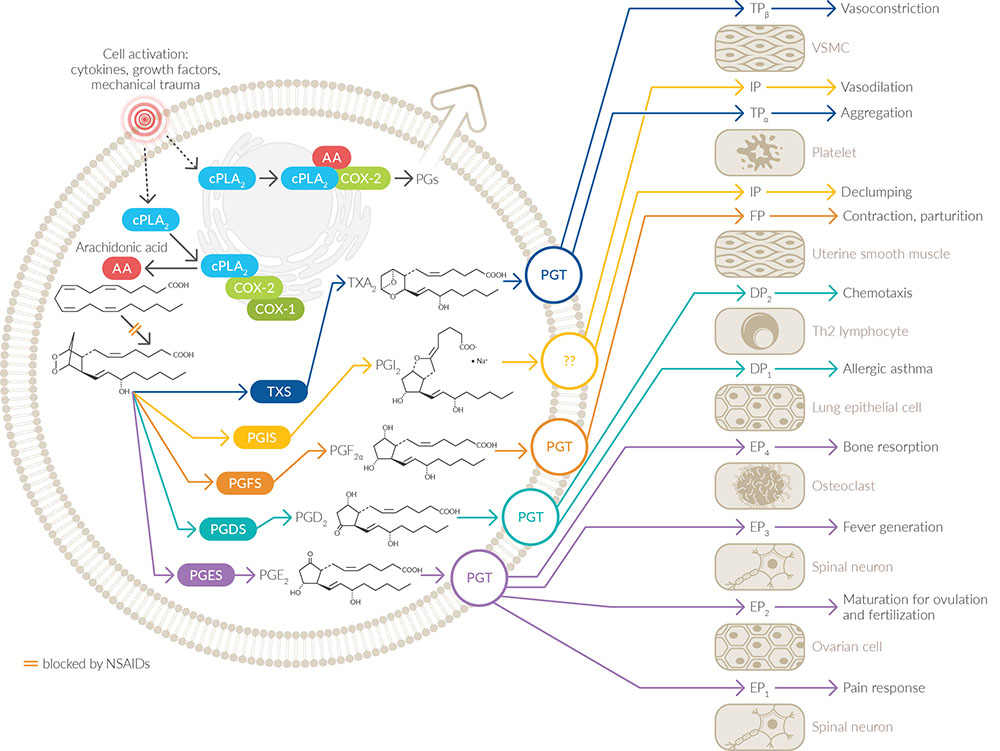
Synthesis and activities of the primary prostaglandins and thromboxane. Image used with permission from Colin Funk.
Original source: Funk, C.D. Science 294(5548), 1871-1875 (2001).
Further metabolism of these initial lipid mediators typically reduces biological activity substantially. The primary route of prostaglandin metabolism is through the 15-hydroxy prostaglandin dehydrogenase pathway. Enzymes in this pathway first oxidize the hydroxyl to a ketone at carbon 15. This is followed by reduction of the double bond at carbons 13 and 14 by 15-ketoprostaglandin Δ13-reductase. Subsequent beta and omega oxidation on the top and bottom side chains, respectively, yields the final metabolic products that can be measured in urine.
Prostaglandin metabolism from source to excretion.
Leukotrienes (LTs) are synthesized in leukocytes through a unique transcellular process in which the precursor LTA4 is made in one cell and transferred to another cell where it is converted to a subsequent product (for simplicity, the entire pathway is depicted in a single cell type below). Cell activation resulting in an increase in intracellular calcium causes translocation of cPLA2 and 5-LO to intracellular membranes where it is joined by 5-LO-activating protein (FLAP). These three proteins work in concert to produce LTA4 from membrane-bound arachidonic acid. LTA4 can then be converted to either LTB4 by the enzyme LTA4 hydrolase, or to LTC4 by LTC4 synthase. Both enzymes are drug targets for the development of anti-inflammatory drugs. The LTs are transported out of the cell by membrane-bound transporters where they then bind to specific GPCRs on adjacent cells to mediate cell- and tissue-specific effects.
Synthesis and activities of leukotrienes. Image used with permission from Colin Funk. Original source: Funk, C.D. Science 294(5548), 1871-1875 (2001).
You May Also Be Interested In
Contact Info
Cayman Chemical1180 East Ellsworth RoadAnn Arbor, Michigan 48108 USAToll Free: (800) 364-9897(USA and Canada only)Fax: (734) 971-3640


