
News & Announcements
The Role of Sulfatides in Disease
Article from 2018-02-01
This article was originally published in the February 2018 edition of Matreya’s Newsletter for Glyco/Sphingolipid Research (PDF).
Sulfatides are 3-sulfated galactosylceramides that are found primarily in the central nervous system and are abundant in the myelin sheath and myelinating cells. Over the last several decades, sulfatides have been linked to many physiological functions and recently there has been a renewed interest in their role in diseases. Sulfatides are highly multifunctional glycolipids involved in the nervous system, diabetes, immune system, hemostasis/thrombosis, and bacterial and viral infection. By understanding the relationship between normal physiological functions of sulfatides and their specific roles in disease, new diagnostic and therapeutic methods can be evaluated.
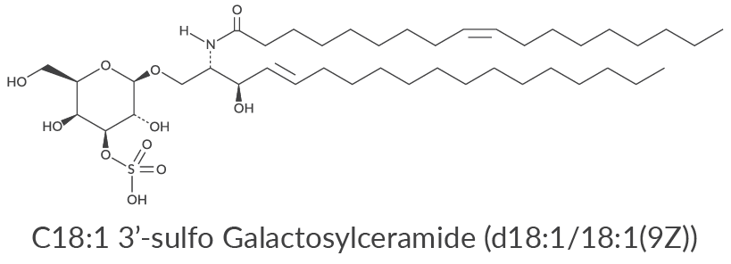
Sulfatides derived from the brain and spinal cord can have saturated, unsaturated, and 2-hydroxy fatty acyl chains, the composition of which is vital to influencing their function. They have demonstrated many critical physiological processes, both inter- and intracellularly, and are involved in numerous diseases such as demyelinating diseases. Various infections, including influenza virus A and tuberculosis, have been shown to be influenced by the actions of sulfatides.1 Recent studies have implicated sulfatides in numerous inflammatory responses, and there has been significant interest in their biological role towards CD1-restricted T cells. An important physiological role of sulfatides is their involvement in hemostasis and thrombosis by forming stable platelet aggregates.2,3 In ovarian cancer, sulfatide levels have been found to be elevated, with the most prevalent species detected via MALDITIMS being d18:1/C16:0, d18:1/C24:1, and d18:1/C24:0.4 This elevation in sulfatide levels can be exploited as an ovarian cancer biomarker and as a possible therapeutic approach against the disease. In hepatic ischemic reperfusion injury, subsets of natural killer T (NKT) cells have opposing roles.5 Type I NKT cells promote injury while sulfatide-reactive type II NKT cells protect against injury. CD1d activation of NKT cells is conserved from mice to humans, so strategies to modify these processes might be developed to treat patients with hepatic reperfusion injury. Abnormal sulfatide metabolism, such as in metachromatic leukodystrophy, can induce cell apoptosis due to endosome-mediated ceramide generation and the accumulation of cytotoxic levels of sulfatides in lysosomes.6 Metachromatic leukodystrophy is an autosomal-recessive lysosomal storage disease caused by mutations in the arylsulfatase A (ARSA) gene, leading to ARSA deficiency and causing sulfatide accumulation. The main symptoms of the disease are progressive demyelination, neurological dysfunction, and reduced life expectancy.7
Due to its prevalence in the myelin sheath of nerves, it is not surprising that sulfatide metabolism has been implicated in many neural degenerative diseases such as Alzheimer's disease and multiple sclerosis. Sulfatide content is found to precipitously drop in Alzheimer's disease, with its concentration in the central nervous system being modulated by apolipoprotein E.8 Multiple sclerosis is a chronic inflammatory disease of the central nervous system where the myelin sheath around nerve fibers becomes the target of an autoimmune attack leading to demyelination, axonal loss, and subsequent progressive functional neurological deficits. The identity of the target antigen of multiple sclerosis has yet to be discovered, but it was recently demonstrated that levels of anti-sulfatide antibodies were significantly higher in multiple sclerosis patients than in controls.9 Sulfatides are also able to activate inflammatory responses as an endogenous stimulator in brain-resident immune cells.10
These sulfatides bind to several human CD1 molecules as well as to mouse CD1d and are recognized by type II NKT cells. Among the sulfatides, cis-tetracosenoyl sulfatide is immunodominant and can either prevent or reverse antigen-induced experimental autoimmune encephalomyelitis in CD1d+/+ mice.11 It therefore may also be able to reverse autoimmune demyelinating diseases in humans.12 It has been demonstrated that the sulfatide presented on the major histocompatibility complex (MHC) class I-like CD1d and CD1c molecules was recognized by Vδ1 lymphocyte cells. The myelin sheath, the target of the autoimmune attack in multiple sclerosis, is a rich source of sulfatides, suggesting that CD1c/d-restricted T cells could be activated by myelin-derived sulfatide and, thereby, could be implicated in the onset of the disease.13 A depletion of different sulfatide species from the earliest stages of multiple sclerosis in both white and gray matter areas of the frontal cortex could be considered a marker of the disease, but may also indicate neurochemical modifications related to its pathogenesis.14 Sulfatides are highly dynamic glycosphingolipids with far-reaching biological processes. A greater understanding of these functions in living systems will lead to the elucidation of associated diseases and the development of therapeutic treatments for various sulfatide-associated diseases.
Available Sulfatides
Sulfatides (bovine) (sodium salt)
3’-sulfo Galactosylsphingosine (ammonium salt)
C2 3'-sulfo Galactosylceramide (d18:1/2:0)
C12 3'-sulfo Galactosylceramide (d18:1/12:0)
C16 3'-sulfo Galactosylceramide (d18:1/16:0)
C17 3'-sulfo Galactosylceramide (d18:1/17:0)
C18 3'-sulfo Galactosylceramide (d18:1/18:0)
C18:1 3'-sulfo Galactosylceramide (d18:1/18:1(9Z))
C18 3'-sulfo Lactosylceramide (d18:1/18:0)
C19 3'-sulfo Galactosylceramide (d18:1/19:0)
C24 3'-sulfo Galactosylceramide (d18:1/24:0)
C24:1 3'-sulfo Galactosylceramide (d18:1/24:1(15Z))
C18 3'-sulfo Galactosylceramide-d3 (d18:1/18:0-d3)
C12 NBD 3'-sulfo Galactosylceramide (d18:1/12:0)
C6 Biotin 3'-sulfo Galactosylceramide (d18:1/6:0)
N-Glycine 3’-sulfo Galactosylsphingosine
You Might Be Interested In…
The Biological Role of Sulfatides
Sulfatides in Alzheimer's Disease and Multiple Sclerosis
Sulfatides in Metachromatic Leukodystrophy
References
1. Suzuki, T., Sometani, A., Yamazaki, Y., et al. Sulphatide binds to human and animal influenza A viruses, and inhibits the viral infection. Biochem. J. 318(Pt 2), 389-393 (1996).
2. Merten, M., Beythien, C., Gutensohn, K., et al. Sulfatides activate platelets through P-selectin and enhance platelet and platelet-leukocyte aggregation. Arterioscler. Thromb. Vasc. Biol. 25(1), 258-263 (2005).
3. Kyogashima, M. The role of sulfatide in thrombogenesis and haemostasis. Arch. Biochem. Biophys. 426(2), 157-162 (2004).
4. Liu, Y., Chen, Y., Momin, A., et al. Elevation of sulfatides in ovarian cancer: An integrated transcriptomic and lipidomic analysis including tissue-imaging mass spectrometry. Mol. Cancer 9, 186 (2010).
5. Arrenberg, P., Maricic, I., and Kumar, V. Sulfatide-mediated activation of type II natural killer T cells prevents hepatic ischemic reperfusion injury in mice. Gastroenterology 140(2), 646-655 (2011).
6. Zeng, Y., Cheng, H., Jiang, X., et al. Endosomes and lysosomes play distinct roles in sulfatide-induced neuroblastoma apoptosis: Potential mechanisms contributing to abnormal sulfatide metabolism in related neuronal diseases. Biochem. J. 410(1), 81-92 (2008).
7. Dehghan Manshadi, M., Kamalidehghan, B., Aryani, O., et al. Four novel ARSA gene mutations with pathogenic impacts on metachromatic leukodystrophy: A bioinformatics approach to predict pathogenic mutations. Ther. Clin. Risk Manag. 13, 725-731 (2017).
8. Zeng, Y. and Han, X. Sulfatides facilitate apolipoprotein E-mediated amyloid-β peptide clearance through an endocytotic pathway. J. Neurochem. 106(3), 1275-1286 (2008).
9. Ilyas, A.A., Chen, Z.W., and Cook, S.D. Antibodies to sulfatide in cerebrospinal fluid of patients with multiple sclerosis. J. Neuroimmunol. 139(1-2), 76-80 (2003).
10. Jeon, S.-B., Yoon, H.J., and Park, S.-H. Sulfatide, a major lipid component of myelin sheath, activates inflammatory responses as an endogenous stimulator in brain-resident immune cells. J. Immunol. 181(11), 8077-8087 (2008).
11. Halder, R.C., Aguilera, A., Maricic, I., et al. Type II NKT cell-mediated anergy induction in type I NKT cells prevents inflammatory liver disease. J. Clin. Invest. 117(8), 2302-2312 (2007).
12. Halder, R.C., Jahng, A., Maricic, I., et al. Mini review: Immune response to myelin-derived sulfatide and CNS-demyelination. Neurochem. Res. 32(2), 257-262 (2007).
13. Singh, A.K., Novakova, L., Axelsson, M., et al. High interferon-γ uniquely in Vδ1 T cells correlates with markers of inflammation and axonal damage in early multiple sclerosis. Front. Immunol. 8, 260 (2017).
14. Gónzalez de San Román, E., Manuel, I., Giralt, M.T., et al. Imaging mass spectrometry (IMS) of cortical lipids from preclinical to severe stages of Alzheimer's disease. Biochim. Biophys. Acta. Biomembr. 1859(9 Pt B), 1604-1614 (2017).
Contact Info
Cayman Chemical1180 East Ellsworth RoadAnn Arbor, Michigan 48108 USAToll Free: (800) 364-9897(USA and Canada only)Fax: (734) 971-3640


