
News & Announcements
Capturing NETosis: Finding the Right Assay Kit for Your Application
Featured Article from 2026-08-31
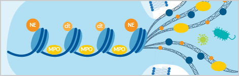
The deployment of neutrophil extracellular traps (NETs) in a process termed NETosis is one of several mechanisms used by neutrophils to defend against pathogens.1,2 During NETosis, decondensed chromatin is extruded into the extracellular environment to form web-like structures of DNA decorated with citrullinated histone H3 (CitH3) and granule proteins such as myeloperoxidase (MPO) and neutrophil elastase (NE).1,3 NETosis can be triggered by stimuli including pathogen-associated and host-derived damage-associated molecular patterns (PAMPs and DAMPs, respectively), as well as pharmacological agents including phorbol 12-myristate 13-acetate (PMA), calcium ionophores (e.g., ionomycin, A23187), and the potassium ionophore nigericin.1,3,4 Both the composition of NETs and the kinetics of their formation are influenced by the stimulus used to induce NETosis.4,5
While NETosis is profoundly useful for its ability to ensnare and neutralize microbial pathogens, it is a double-edged sword in that aberrant NETosis or defects in NET clearance have been implicated in numerous inflammatory disorders and autoimmune diseases, including systemic lupus erythematosus (SLE), psoriasis, and rheumatoid arthritis.3,6,7
Because no single assay captures every aspect of NETosis, Cayman Chemical has developed a suite of complementary tools to support NET detection, quantification, and visualization.
Tools to Study NETosis
Explore the selection guide below to learn more about our industry-leading solutions and determine the best assay(s) for your application.
| Best for | Product Name | Item No. |
| Detecting NETs in biological matrices | NET Fragment Assay Kit (MPO-DNA) | 501330 |
| Citrullinated Histone H3 (Clone 11D3) ELISA Kit | 501620 | |
| Evaluating stimulators and inhibitors of NETosis in vitro | NETosis Assay Kit | 601010 |
| NETosis visualization and kinetic studies | NETosis Imaging Assay Kit | 601750 |
Best for Detecting NETs in Biological Matrices: Citrullinated Histone H3 (Clone 11D3) ELISA Kit & NET Fragment Assay Kit (MPO-DNA)
Given the complexity and heterogeneity of NETs, there is as yet no universally accepted NET biomarker. Therefore, current best practices recommend combining detection of multiple NET markers for more accurate analysis.
Cayman offers convenient assay kits to detect CitH3 and MPO-DNA complexes (i.e., NET fragments), two of the most widely used NET-associated biomarkers, in biological matrices. Our Citrullinated Histone H3 (Clone 11D3) ELISA Kit has been cited in approximately 100 scientific publications, and our new NET Fragment Assay Kit (MPO-DNA) provides a solution to the current lack of standardization and commercially available options to study this key biomarker.
| Product Name | NET Fragment Assay Kit (MPO-DNA) | Citrullinated Histone H3 (Clone 11D3) ELISA Kit | |
| Item No. | 501330 | 501620 | |
| Biomarker Detected | Intact MPO-DNA complexes (i.e., NET fragments) | CitH3 | |
| Assay Format | Immunometric (i.e., sandwich) assay | Immunometric (i.e., sandwich) assay | |
| Quantitative or Qualitative? | Qualitative | Quantitative | |
| Assay Range | N/A | 0.15-10 ng/ml | |
| Validated Matrices | |||
Human plasma | ✅ (EDTA and citrate) | ✅ | |
Human serum | X | ✅ | |
Cell culture supernatants | ✅ | ✅ | |
Cell lysates | X | ✅ | |
| Run Time | Under 4 hours | Under 4 hours | |
| Samples | Up to 46 samples in duplicate or 30 samples in triplicate | Assay 36 samples in duplicate or 24 samples in triplicate | |
| Readout | Colorimetric (450 nm) | Colorimetric (450 nm) | |
| Best For | Detecting MPO-DNA complexes in plasma and supernatants | Quantification of CitH3 across multiple sample types | |
Briefly, the NET Fragment Assay Kit (MPO-DNA) uses an immunometric (i.e., sandwich) assay format with an anti-MPO capture antibody and anti-DNA HRP conjugate detection antibody to detect intact MPO-DNA complexes. The Citrullinated Histone H3 (Clone 11D3) ELISA Kit also uses a "sandwich" assay format, but with an anti-CitH3 capture antibody and anti-histone H3 HRP conjugate detection antibody. In both assays, the HRP substrate TMB is added to the wells, yielding a reaction product that can be measured at 450 nm.
View complete assay kit details and protocol for the NET Fragment Assay Kit (MPO-DNA) in the kit booklet (PDF) and performance and validation data in the validation data PDF. Full assay kit details, protocol, and validation for the Citrullinated Histone H3 (Clone 11D3) ELISA Kit can be found in the kit booklet (PDF).
Are My Samples Compatible?The NETosis assay kits discussed in this article have all been validated in human samples, however they may also be compatible with additional species or sample types. For questions regarding species compatibility or validation of additional sample matrices, contact our Technical Support team. |
Best for In Vitro NETosis Studies: NETosis Assay Kit
Cayman's NETosis Assay Kit allows for in vitro induction and detection of NETs via measurement of NET-derived neutrophil elastase (NE) activity and can be used for quantitative evaluation of stimulators and inhibitors of NETosis.
| Product Name | NETosis Assay Kit |
| Item No. | 601010 |
| Biomarker Detected | NET-derived neutrophil elastase activity |
| Assay Format | Cell-based enzymatic assay |
| Quantitative or Qualitative? | Quantitative |
| Validated Matrices | Primary human neutrophils |
| Samples | Up to 80 individual samples or 24 samples in duplicate |
| Readout | Colorimetric (405 nm) |
In this assay, primary neutrophils are incubated with chosen stimuli (both PMA and A23187 are included in the kit) to induce NETosis (Figure 1). Following generation of NETs, soluble NE is removed in a wash step, leaving intact NETs and associated NE in the wells. Next, S7 nuclease is added to the wells to digest NET DNA, releasing NET components including NE into the supernatant, which is collected and added to a substrate that is selectively cleaved by NE to yield a product that absorbs light at 405 nm.
Figure 1. Step-by-step diagram of NET formation and analysis.
View complete assay kit details, protocol, and validation data in the kit booklet (PDF).
Need Help Isolating Neutrophils?Our Neutrophil (human) Isolation Kit allows for isolation of neutrophils from human whole blood, and our Neutrophil (mouse) Isolation Kit offers a streamlined method to isolate mouse neutrophils from peritoneal lavage or bone marrow. |
Best for NETosis Visualization & Kinetic Studies: NETosis Imaging Assay Kit
Cayman's NETosis Imaging Assay Kit provides a convenient method to visualize NETosis and study its kinetics.
| Product Name | NETosis Imaging Assay Kit |
| Item No. | 601750 |
| Biomarker Detected | Extruded DNA |
| Assay Format | Cell-based fluorometric assay |
| Quantitative or Qualitative? | Qualitative |
| Validated Matrices | Primary human neutrophils |
| Samples | Sufficient reagents for two 96-well plates |
| Readout | Fluorescence (ex/em = 503/526 and 622/645 nm for green and red dyes, respectively) |
In this assay, isolated neutrophils are treated with a cell-permeable DNA dye, which enables visualization of the dynamic nuclear changes during NETosis, as well as a cell-impermeable dye, which detects extruded DNA. NETosis is induced by addition of chosen stimuli (both PMA and A23187 are included in the kit) and monitored using brightfield and fluorescence microscopy (Figure 2). Combining brightfield and fluorescence imaging in a high-content platform allows for examination of the morphological changes associated with NETosis.
Figure 2. Monitoring the morphology of primary human neutrophils undergoing NETosis. DNA extruded during NETosis is detected by Extracellular Nuclear Green™ Reagent in neutrophils stimulated with PMA or nigericin. Intact nuclei are counterstained with Permeable Nuclear Red™ Reagent. Timed images (every hour) were generated with BioTek®’s Cytation™ 5 Cell Imaging Multi-Mode Reader using a 20X objective and GFP and Cy5 LED filter combinations with brightfield. Scale bars are 30 µm, and filled and empty arrowheads follow individual cells undergoing NETosis.
Along with the necessary reagents for DNA staining and NET induction, this assay kit also includes reagents to isolate neutrophils from human blood.
View full kit details, protocol, and validation data in the kit booklet (PDF).
The Cayman Advantage
At Cayman, we have an exceptional understanding of assay development, validation, and performance. Driven by the growing interest in the roles of NETosis in physiological and pathological contexts, our scientists put their assay development expertise to work to develop a comprehensive NETosis toolkit that meets researchers' needs for optimized, validated tools to study different aspects of this process.
The NETosis assay kits featured above are part of our extensive and trusted line of NETosis research tools, including inhibitors and inducers of NETosis as well as proteins, antibodies, and additional assay kits to study neutrophil biology.
Our scientists also offer support beyond the sale, with expert technical support readily available to assist you with any troubleshooting needs.
You May Also Be Interested In
References
1. Poli, V. and Zanoni, I. Neutrophil intrinsic and extrinsic regulation of NETosis in health and disease. Trends Microbiol. 31(3), 280-293 (2023).
2. Thiam, H.R., Wong, S.L., Wagner, D.D., et al. Cellular mechanisms of NETosis. Annu. Rev. Cell Dev. Biol. 36, 191-218 (2020).
3. Gillot, C., Bouarroudj, H., Decarpentrie, J., et al. Techniques for measuring NETosis: A critical literature review and outlook for standardization. Thromb. Res. 262, 109697 (2026).
4. Kenny, E.F., Herzig, A., Krüger, R., et al. Diverse stimuli engage different neutrophil extracellular trap pathways. eLife 6, e24437 (2017).
5. Neeli, I. and Radic, M. Opposition between PKC isoforms regulates histone deimination and neutrophil extracellular chromatin release. Front. Immunol. 4, 38 (2013).
6. Vorobjeva, N.V. and Pinegin, B.V. Neutrophil extracellular traps: Mechanisms of formation and role in health and disease. Biochemistry (Mosc.) 79(12), 1286-1296 (2014).
7. Pinegin, B., Vorobjeva, N., and Pinegin, V. Neutrophil extracellular traps and their role in the development of chronic inflammation and autoimmunity. Autoimmun. Rev. 14(7), 633-640 (2015).
Contact Info
Cayman Chemical1180 East Ellsworth RoadAnn Arbor, Michigan 48108 USAToll Free: (800) 364-9897(USA and Canada only)Fax: (734) 971-3640


