
News & Announcements
An Introduction to Lipid Nanoparticle Formulation: Basic Concepts & Preparation Procedures
Article from 2022-02-23
By Christine R. Barillas, Ph.D. and Melissa A. Parsey, Ph.D. - Technical Writers, Cayman Chemical
The success of mRNA-based COVID-19 vaccines could not have been possible without decades of research on lipid-based drug delivery (LBDD). LBDD systems are highly versatile and have been used to deliver various bioactive molecules to targeted cells and tissues. LBDD has several advantages over conventional drug delivery methods, including increased drug stability, bioavailability, and distribution. Lipid nanoparticles (LNPs) are a significant advancement for the delivery of nucleic acid-based therapeutics. Nucleic acids encapsulated within LNPs are protected from enzymatic degradation during the delivery process and are efficiently delivered to cells, where the therapeutic cargo is released.
Here, we introduce the basic principles of common LBDD systems and the cargoes they deliver, with an emphasis on LNPs. This background provides the foundation for understanding why and how LNPs deliver nucleic acid therapies. We then cover the basic concepts and procedures for the preparation of LNPs, including:
Key Definitions
This glossary is intended to introduce you to terms commonly used to describe LBDD systems. Here, we summarize the structure and type of cargo encapsulated in these different systems.
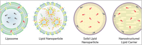
- Lipid nanoparticles (LNPs): a lipid shell surrounding an internal core composed of reverse micelles that encapsulate and deliver nucleic acids, like siRNA and mRNA, and plasmid DNA (pDNA) (Table 1).
- Liposomes: contain one or more lipid bilayers and an aqueous core. They are further classified by lamellarity and size. Liposomes are used for hydrophobic and/or hydrophilic small molecules.
- Solid lipid nanoparticles (SLNs): a surfactant shell surrounding a core matrix composed of solid lipids. They are used for hydrophobic and/or hydrophilic cargo.
- Nanostructured lipid carriers (NLCs): a surfactant shell surrounding a core matrix composed of solid and liquid lipids. They are used for hydrophobic and/or hydrophilic cargo.
- Micelles: self-assemblies of lipid monolayers in aqueous solutions. They have a hydrophobic core, where the phospholipid tails are oriented towards the interior, and can be used for the encapsulation of small hydrophobic cargo.
- Reverse micelles: an inverted structure compared with traditional micelles. They form a hydrophilic core, with the phospholipid tails oriented towards the exterior, and can be used for the encapsulation of small hydrophilic cargo, like nucleic acids in LNPs.
Table 1: LNPs compared to other LBDD systems
| | Lipid Nanoparticle | Liposome | Solid Lipid Nanoparticle | Nanostructured Lipid Carrier |
| | ||||
| Lipid Shell | Monolayer | Bilayer | Surfactant | Surfactant |
| Internal Core | Reverse micelles | Aqueous | Solid lipids | Solid and liquid lipids |
| Cargo | Nucleic acids | Hydrophobic and/or hydrophilic small molecules | Hydrophobic and/or hydrophilic small molecules | Hydrophobic and/or hydrophilic small molecules |
| Size | ~50-150 nm | ~50-1,000 nm | ~40-1,000 nm | ~40-1,000 nm |
Structural Components
LNPs are composed of ionizable cationic lipids, helper lipids, which include glycerophospholipids, sterol lipids, and PEGylated lipids to protect nucleic acids, which are contained within an aqueous phase. Many of the same structural components used in LNPs are components of other LBDD particles. Lipids and molecules that contain them, like surfactants, can be used to tailor the behavior and properties of LBDD particles. Therefore, the selection of structural components is important to consider during LBDD particle formulation.
Ionizable cationic lipids circumvent the untoward cytotoxicity associated with cationic lipids. These lipids possess a transient cationic charge that is acquired at low pH (typically <7), forming reverse micelles that encapsulate nucleic acids in the LNP core. As these lipids have near-neutral charge at physiological pH, they deliver nucleic acid cargo without cytotoxicity. There are five major structural classes of ionizable cationic lipids that have varying effects on RNA delivery and toxicity: unsaturated, multi-tail, branched-tail, polymeric, and biodegradable ionizable lipids. Select ionizable cationic lipids available from Cayman and their pKa values can be found at the end of this article. |
Glycerophospholipids, colloquially known as helper lipids, are a class of phospholipid that contains a hydrophilic head group and two hydrophobic fatty acyl tails attached to a glycerol backbone. The hydrophilic head determines the charge of the LBDD particle, which can be neutral, anionic (negative), or cationic (positive).
Neutral phospholipids improve the efficacy of membrane fusion and can distribute or modify the net surface charge of the lipid particle. The phospholipid head groups with an overall neutral charge are phosphatidylcholine (PC) and phosphatidylethanolamine (PE). |
Sterol Lipids, such as cholesterol, are used to fill lipid membrane packing defects and provide structural integrity. They also aid in membrane fusion of the LNP and target cell. |
PEGylated Lipids prevent serum protein adsorption, inhibiting uptake by the mononuclear phagocyte system (MPS), a major obstacle in the delivery of LBDD systems. Some PEGylated lipids also contain terminal functional groups, such as amine or maleimide, which can be used to conjugate other molecules that improve cellular targeting and uptake. |
Aqueous Phase
The aqueous phase solubilizes hydrophilic molecules, like nucleic acids in LNPs and hydrophilic drugs in liposomes, whereas hydrophobic cargo can be solubilized in the lipid phase. |
Surfactants
Surfactants are a major component of the lipid shell of SLNs and NLCs. They reduce the interfacial tension between the lipophilic core and aqueous phase due to their amphipathic nature and improve stability during storage. Lecithin, which contains phosphatidylcholines, is one surfactant that has been used in SLNs and NLCs. |
Solid Lipids
Solid lipids, colloquially known as fats, are solid at ambient temperature and used in the preparation of both SLNs and NLCs. Solid lipids used to prepare SLNs or NLCs are typically saturated and include glycerolipids, such as triacylglycerols, and fatty acids, including stearic acid, as well as fatty alcohols and fatty esters. |
Liquid Lipids
Liquid lipids, also referred to as oils, are fluid at ambient temperature. NLCs are formulated with a mixture of solid and liquid lipids, which increases drug loading capacity and prevents drug leakage. Liquid lipids used in NLCs are typically unsaturated and include oleic acid, α-tocopherol, and squalene. |
Read our article The Heads and Tails of Lipid-Based Drug Delivery to dive deeper into the biophysical properties of lipids used in LBDD systems. |
Cargo
LNPs are superior for the encapsulation of nucleic acids, whereas other LBDD systems are preferable for the delivery of small molecule inhibitors or lipids. The requisite LBDD system for your application depends on the cargo, and the localization of the cargo within the LBDD particle depends on its physicochemical properties.
Nucleic Acids
mRNA, siRNA, and pDNA are common nucleic acid cargo. Nucleic acids are negatively charged and best encapsulated within LNPs using ionizable cationic lipids. mRNA-containing LNPs are the basis for most COVID-19 vaccines, and LNPs containing transthyretin-targeting siRNA have been developed for the treatment of hereditary amyloidogenic transthyretin (ATTRv) amyloidosis. |
Small Molecules
Hydrophobic and/or hydrophilic small molecules can be solubilized in the aqueous or lipophilic compartments, respectively, of liposomes, SLNs, and NLCs.
Hydrophobic drugs are dispersed throughout the lipophilic compartments of LBDD systems. Amphotericin B, an antifungal agent, and verteporfin, a photosensitizing agent, are examples of FDA-approved hydrophobic drugs that have been formulated in liposomes. | |
Hydrophilic drugs are solubilized in the aqueous compartments of LBDD systems. Doxorubicin, an antitumor antibiotic, is a hydrophilic drug. Doxil® is a form of doxorubicin encapsulated in liposomes and was the first LBDD formulation to be approved by the FDA. |
LNP Design
Several factors should be considered when selecting lipids and how they are formulated into LNPs. Below, we outline these factors and how each of them influences the physiological behavior of LNP particles.
The lipid molar ratio determines the lipid composition of the particles and influences their size, polydispersity, and efficacy. We recommend starting with a literature review to identify lipid molar ratios that have been previously developed for similar applications. Lipid molar ratios for FDA-approved LNPs are as follows (Table 2): |
Table 2. Lipid molar ratios for LNPs in FDA-approved agents
| | Patisiran | BNT162b2 | mRNA-1273 |
| Cargo | siRNA | mRNA | mRNA |
| Ionizable Cationic Lipid | DLin-MC3-DMA | ALC-0315 | SM-102 |
| Neutral Phospholipid | 1,2-DSPC | 1,2-DSPC | 1,2-DSPC |
| Sterol Lipid | Cholesterol | Cholesterol | Cholesterol |
| PEGylated Lipid | C-DMG-PEG(2000) | ALC-0159 | DMG-PEG(2000) |
| Lipid Molar Ratioa | 50:10:38.5:1.5 | 46.3:9.4:42.7:1.6 | 50:10:38.5:1.5 |
aIonizable cationic lipid:neutral phospholipid:cholesterol:PEGylated lipid
The lipid:nucleic acidweight ratio influences the encapsulation efficiency. Most LNPs are formulated with a lipid:oligonucleotide weight ratio of 10-30:1. | |
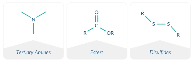
The ionizable lipid nitrogen:nucleic acid phosphate (N:P) molar ratio represents the charge balance between the cationic tertiary amine of the ionizable cationic lipid and the anionic phosphate group in the nucleic acid backbone. This property is the basis for the complexation of ionizable cationic lipids with nucleic acids. LNPs commonly have an N:P ratio around six. | |
The lipid acid dissociation constant (lipid pKa) is the pH at which the ionized and nonionized forms of a lipid exist in equal concentrations. Lipid pKa impacts the LNP encapsulation efficiency, efficacy, delivery, and toxicity. For RNA delivery, the lipid pKa generally ranges from 6-7. | |
Three important parameters for an aqueous buffer are its composition, ionic strength, and pH. Buffers stabilize nucleic acids in solution, and ionizable cationic lipids become protonated and positively charged in the acidic aqueous buffer upon mixing. Commonly used buffers in LNP preparations are 25-50 mM sodium acetate or sodium citrate, pH 4-5. After preparation, LNPs are dialyzed into a neutral buffer, such as PBS, pH 7.4, for storage and use. | |
The particle size alters the pharmacokinetics of the administered particle. Smaller particles typically have longer circulation half-lives, as they evade elimination by the MPS. Particles less than 100 nm can easily pass through fenestrated endothelium to penetrate target tissues. The particle size is dependent on the preparation method. Depending on the LNP preparation method, extrusion can be used to achieve smaller, more uniform particle sizes. | |
The two most commonly used routes of administration for LNPs are intravenous and intramuscular injection. Of note, formulations optimized for a given route of administration are generally not applicable for other routes of administration. Intravenously administered LNPs with net positive, neutral, and negative charges can be targeted to the lungs, liver, and spleen, respectively. The inclusion of cholesterol or PEGylated lipids in the formulation, as well as increasing the LNP size, increases distribution to the spleen. Intramuscular administration is commonly used for vaccines, as it facilitates lymph node targeting and activation of the immune response. When a vaccine is administered, antigen-presenting cells (APCs), like macrophages and dendritic cells, are recruited to the delivery site, where they can encounter vaccine antigens. They then migrate to lymph nodes where they stimulate adaptive immune responses. | |
The preparation method determines the properties of LNPs, including size, homogeneity, and encapsulation efficiency. When selecting a preparation method, cost, scalability, reproducibility, and time commitment should also be considered. |
Procedures
In the sections below, we give a series of a simple protocols to follow for producing LNPs. The entire range of the LNP life cycle is covered, starting with LNP preparation at the bench and ending with how to use and what to expect when using LNPs for your in vitro or in vivo experiments.
LNP Preparation
Before beginning, ensure that all supplies, reagents, and working environments are RNase-free. siRNA and mRNA are chemically labile to RNases, which are enzymes that degrade RNA-based nucleic acids. The steps in LNP formation are summarized in Figure 1.
Figure 1. LNP preparation workflow.
Mixing
LNPs are prepared by mixing an ethanolic lipid mixture with an acidic aqueous buffer containing nucleic acids (Figure 2). A 1:3 ratio of ethanolic lipid mixture to aqueous buffer is generally used. Several methods are suitable for laboratory-scale, small-volume LNP production. Two of these methods, suitable for a range of specialized-to-basic equipment, are compared briefly below and in Table 3.
Figure 2. Schematic of oligonucleotide-containing LNP formation.
Microfluidic Mixing Devices: Automated microfluidic devices or microfluidic chips are fast and efficient methods to prepare LNPs. These devices enable rapid mixing in a highly controllable, reproducible manner that achieves homogeneous LNPs and high encapsulation efficiency. In these devices, individual streams of the ethanolic lipid mixture and aqueous oligonucleotide solution are rapidly combined. LNPs form as the two streams mix and are collected into a single collection tube. Parameters such as the flow rate ratio (FRR) and total flow rate (TFR) can be altered to fine-tune LNPs. | |
Hand-mixing: Transfer the ethanolic lipid mixture into the aqueous acidic oligonucleotide solution and mix for 15 seconds by rapid pipetting. Leave the mixture undisturbed for 10 minutes. Hand-mixing of LNPs results in heterogeneous LNPs with low encapsulation efficiency and can yield variable results. |
Table 3. Feature comparison of LNP preparation methods
| Cost | Scalability | Encapsulation Efficiency | Reproducibility | Polydispersity Index | |
|---|---|---|---|---|---|
Microfluidic Mixing Devices | High | High | High | High | Low |
Hand-mixing | Low | Low | Low | Low | High |
Final Preparation
The final preparation of LNPs is performed after they have been formed during the mixing step. The steps that follow help ensure that the LNPs are homogeneous, stable during storage and use, and free of any residual chemical or biological contaminants.
Extrusion | |
Dialysis | |
Filter-sterilize |
Stability & Storage
After LNPs have been prepared, they may either be used immediately or stored for later use. Below, we discuss factors that can compromise LNP integrity during storage and provide tips on how to limit storage instability.
Physical stability describes the structural integrity of LNPs during storage. Particle fusion or aggregation and leakage of encapsulated cargo are examples of physical instability.
🗸 Ensure size distribution remains small and homogeneous:
|
Chemical stability defines the resistance of LNP lipid and cargo components to modifications in their molecular structure. Hydrolysis, oxidation, and transesterification can lead to nucleic acid and lipid degradation or the formation of lipid-nucleic acid adducts and loss of efficacy.
🗸 Limit cargo degradation:
|
Biological stability relates to the capacity of LNPs to avoid early degradation. Factors that contribute to biological stability include lipid composition, particle size, and surface charge.
🗸 Reduce serum protein opsonization:
|
Storage is a critical parameter in the stability of LNP formulations.
Generally, LNPs may be stored at 4°C for up to 1 week or, for long-term storage, lyophilized and held at -80°C. Storage temperature, buffers, and pH may need to be optimized. The inclusion of cryoprotectants is recommended when freezing with or without lyophilization. |
Characterization
Characterization of LNP attributes prior to in vitro or in vivo use is critical for reproducibility.
The LNP size describes the average diameter of LNPs and influences their biodistribution and cellular uptake. The polydispersity index (PDI) is a measure of the LNP size distribution. Homogeneous, uniformly sized samples have small PDIs, and samples with heterogeneous size distributions have large PDIs. The LNP size and PDI of an LNP preparation can be reduced by optimizing lipid components, increasing the mixing rate, selecting a different preparation method, or by adding an extrusion step. These attributes can be measured by dynamic light scattering (DLS) using a specialized instrument. | |
Zeta potential is the electrostatic potential surrounding the LNP. In general, a near-neutral zeta potential is desirable. Anionic LNPs may be electrostatically repelled from negatively charged plasma membranes, and cationic LNPs can be cytotoxic. The zeta potential can be adjusted by altering the N:P. Zeta potential is measured by a specialized instrument. | |
Encapsulation efficiency is the final amount of nucleic acid contained within the LNP compared to nucleic acid not encapsulated within the LNP. Microfluidic mixing yields the highest encapsulation efficiencies. Encapsulation efficiency can be measured using nucleic acid-binding fluorescent dyes in the presence and absence of detergent, such as 1% Triton X-100. This technique can also serve to report the encapsulated nucleic acid concentration. | |
Lipid and cargo integrity are essential for the efficacy and stability of LNPs. Refer to the storage and stability section below for more information. Our Chemical Synthesis team offers lipid characterization services. | |
The apparent/global pKa of formulated LNPs determines the effect of the ionizable lipid on the apparent pKa of LNPs. This parameter influences the LNP ionization and surface charge, stability, potency, and toxicity. Cayman's LipidLaunch™ LNP Apparent pKa Assay Kit (TNS Method) contains reagents to determine the ionizable lipid effect on the apparent pKa of LNPs. |
Assessment
LNPs are internalized by target cells via endocytosis. Endosomal escape is the process in which the LNP cargo is delivered to the cytosol. Ionizable cationic lipids become positively charged in the acidic environment of the endosomal lumen, which disrupts the negatively charged endosomal membrane and promotes release of the encapsulated nucleic acid cargo into the cytosol.
Cellular models and simple molecular biology techniques can be used to test the efficacy of LNPs in vitro. Measurement of knockdown or expression of the gene or protein of interest can be accomplished via qPCR or Western blot, respectively. Cell-based reporter or luciferase assays are also used to determine LNP efficacy. | |
Animal models provide valuable insight into the efficacy of LNP-based therapeutics in vivo. Protein expression of nucleic acid products in vivo follows a target-dependent time course. Nucleic acids encoding functional proteins produce changes in protein concentrations within hours, whereas those designed to elicit an antibody response can occur between several days to a couple of weeks. It is often necessary to use repeated dosing regimens to achieve sustained protein expression. ELISAs and multiplexed assays can be used to measure target protein responses, and flow cytometry can determine changes in cellular phenotype. | |
The fate of LNPs after administration depends on the lipid composition, LNP design, and the route of administration. Lipids used in LNPs are detectable in the tissues that LNPs are distributed to after administration. These lipids are biocompatible and rapidly degraded, and they are generally eliminated within 24 to 48 hours after administration. To determine lipid tissue concentrations, mass spectrometry-based approaches can be used. Our Analytical Chemistry team can assist you in the detection of lipids in various tissues and sample matrices. |
Available from Cayman
LBDD systems are at the forefront of gene therapy, vaccine formulations, and targeted drug delivery. The use of high-quality lipids is critical for the success of an LBDD system. Cayman has decades of experience in lipid synthesis and assay development. Our extensive catalog contains over 400 lipids that can be used to form LBDD particles, as well as LipidLaunch™ research-ready LNPs and reagent kits for any step of LNP discovery.

LIPID NANOPARTICLE DEVELOPMENT
From Cayman Services
- End-to-end solutions for LNP-based drug discovery - all from a single-source partner
- Formulation, characterization, & optimization services
- Custom lipid synthesis (CGMP Available)
- High-Purity lipid components
- Ionizable Cationic Lipids
| Structure | Item Name | CAS No. | Item No. | pKa Value of Tertiary Amine |
| | SM-102 | 2089251-47-6 | 33474 | 6.68 |
| | ALC-0315 | 2036272-55-4 | 34337 | 6.09 |
| | Lipid 5 | 2089251-33-0 | 34372 | 6.56 |
| | Lipid 29 | 2244716-55-8 | 35337 | 6.91 |
| | L-319 | 1351586-50-9 | 35051 | 6.38 |
| | DODAP | 127512-29-2 | 25726 | 5.59 |
| | DLin-MC3-DMA | 1224606-06-7 | 34364 | 6.44 |
| | DLin-KC2-DMA | 1190197-97-7 | 34363 | 6.70 |
- Tools to Measure LNP-induced Immune Responses
You May Also Be Interested In
 | |
 Tuning Ionizable Cationic Lipid Design for Efficacy and Safety | |
| Lipid Nanoparticle Development Services |  |
Suggested Reading
Belliveau, N.M., Huft, J., Lin, P.J., et al. Microfluidic synthesis of highly potent limit-size lipid nanoparticles for in vivo delivery of siRNA. Mol. Ther. Nucleic Acids 1(8), e37 (2012).
Beltrán-Gracia, E., López-Camacho, A., Higuera-Ciapara, I., et al. Nanomedicine review: Clinical developments in liposomal applications. Cancer Nanotechnol. 10, 11 (2019).
Duong, V.-A., Nguyen, T.-T.-L., and Maeng, H.-J. Preparation of solid lipid nanoparticles and nanostructured lipid carriers for drug delivery and the effects of preparation parameters of solvent injection method. Molecules 25(20), 4781 (2020).
Eygeris, Y., Gupta, M., Kim, J., et al. Chemistry of lipid nanoparticles for RNA delivery. ACC Chem. Res. 55(1), 2-12 (2022).
Fan, Y., Marioli, M., and Zhang, K. Analytical characterization of liposomes and other lipid nanoparticles for drug delivery. J. Pharm. Biomed. Anal. 192, 113642 (2021).
Hua, S. and Wu, S.Y. The use of lipid-based nanocarriers for targeted pain therapies. Front. Pharmacol. 4, 143 (2013).
Khalil, I.A., Younis, M.A., Kimura, S., et al. Lipid nanoparticles for cell-specific in vivo targeted delivery of nucleic acids. Biol. Pharm. Bull. 43(4), 584-595 (2020).
Lin, P.-C., Lin, S., Wang, P.C., et al. Techniques for physicochemical characterization of nanomaterials. Biotechnol. Adv. 32(4), 711-726 (2014).
Packer, M., Gyawali, D., Yerabolu, R., et al. A novel mechanism for the loss of mRNA activity in lipid nanoparticle delivery systems. Nat. Commun. 12(1), 6777 (2021).
Schoenmaker, L., Witzigmann, D., Kulkarni, J.A., et al. mRNA-lipid nanoparticle COVID-19 vaccines: Structure and stability. Int. J. Pharm. 601, 120586 (2021).
Subramaniam, B., Siddik, Z.H., and Nagoor, N.H. Optimization of nanostructured lipid carriers: Understanding the types, designs, and parameters in the process of formulations. J. Nanopart. Res. 22, 141 (2020).
Tan, J.Y.B, Yoon, B.K., Cho, N.-J., et al. Lipid nanoparticle technology for delivering biologically active fatty acids and monoglycerides. Int. J. Mol. Sci. 22(18), 9664 (2021).
Tenchov, R., Bird, R., Curtze, A.E., et al. Lipid nanoparticles─from liposomes to mRNA vaccine delivery, a landscape of research diversity and advancement. ACS Nano. 15(11), 16982-17015 (2021).
Terada, T., Kulkarni, J.A., Huynh, A., et al. Characterization of lipid nanoparticles containing ionizable cationic lipids using design-of-experiments approach. Langmuir 37(3), 1120-1128 (2021).
Contact Info
Cayman Chemical1180 East Ellsworth RoadAnn Arbor, Michigan 48108 USAToll Free: (800) 364-9897(USA and Canada only)Fax: (734) 971-3640


