
News & Announcements
Deuterated, Odd-Chain, & Fluorescent Standards for Sphingolipidomics
Deuterated, Odd-Chain, & Fluorescent Standards for Sphingolipidomics
Article from 2013-10-01
This article was originally published in the October 2013 edition of Matreya’s Newsletter for Glyco/Sphingolipid Research (PDF).
Over the past decade sphingolipids have gained enormous recognition as vital and complex components of biological systems. For many years, lipids in general, and sphingolipids in particular, have received far less attention than their critical functions deserve. Many reasons contributed to this oversight including the difficulty of extraction and analysis as well as their tremendous diversity in structure and function.1,2 Another historical problem was the very limited availability of appropriate natural and synthetic sphingolipid standards. Fortunately, there has been a welcome advance in making standards available, which has greatly accelerated the analytical field known as sphingolipidomics, leading to our enhanced understanding and classification of sphingolipids.
Sphingolipidomics is the study of the individual sphingolipid species and its metabolism within a given biological system using mass spectrometry. It began to appear as a distinct subfield of the greater lipidomics discipline around 2005.3,4 There are tens of thousands of possible sphingolipids that vary in their polar head groups, acyl chains, and sphingoid bases. The metabolic pathway of these sphingolipids has been extensively studied to understand and treat diseases related to sphingolipids. Many of these sphingolipids are present in only picomole to nanomole amounts, making detection difficult. However, with the incorporation of soft ionization techniques in mass spectrometry, the detection of very small amounts of sphingolipids is possible. Most sphingolipidomics studies use either liquid chromatography (LC)- or shotgun-based mass spectrometry approaches.1 With both approaches, internal standards are used to correct for sample extraction efficiency and instrumentation variability. While adding an internal standard for each individual sphingolipid detected with either of these methods would be ideal, this is impractical due to the vast number of possible sphingolipids in a sample. Thus, internal standards for each class of sphingolipid expected to be found in a sample are used. Appropriate sphingolipidomics internal standards include sphingolipids that are modified on either the oligosaccharide head, ceramide acyl chain, or the sphingosine backbone. These standards are usually stable isotope labeled, unusual in chain length, or fluorescently tagged.
Stable Isotope-Labeled Standards
The most preferred internal standards for lipidomics studies are stable isotope-labeled standards. These standards can be detected by mass spectrometry, while demonstrating nearly identical physical properties compared to natural sphingolipids. This is very important to ensure similar extraction properties between the analytes and the internal standards.1 Most commonly, deuterium or 13C atoms are introduced in the N-acyl chain of the ceramide. The label can also be introduced into the sphingosine tail or oligosaccharide head group, where it is typically more stable.
Stable Isotope-Labeled Sphingolipids
C18 Ceramide-d3 (d18:1/18:0-d3)
C18 dihydro Ceramide-d3 (d18:0/18:0-d3)
C16 Sphingomyelin-13C (d18:1/16:0-13C)
C18 Ceramide-1-phosphate-d3 (d18:1/18:0-d3)
N-Glycine Galactosylsphingosine (d18:1)
C18 Galactosylceramide-d35 (d18:1/18:0-d35)
C18 3'-sulfo Galactosylceramide-d3 (d18:1/18:0-d3)
13C6 Glucosylsphingosine (d18:1)
C16 Glucosylceramide-d3 (d18:1/16:0-d3)
C16 Globotriaosylceramide-d9 (d18:1/16:0-d9)
C18 Globotriaosylceramide-d3 (d18:1/18:0-d3)
C16 Lactosylceramide-d3 (d18:1/16:0-d3)
C16 Ganglioside GM1-d9 (d18:1/16:0-d9) (ammonium salt)
C16 Ganglioside GM2-d9 (d18:1/16:0-d9) (ammonium salt)
C16 Ganglioside GM3-d9 (d18:1/16:0-d9) (ammonium salt)
C18 Ganglioside GM1-d3 (d18:1/18:0-d3) (ammonium salt)
C18 Ganglioside GM2-d3 (d18:1/18:0-d3) (ammonium salt)
C18 Ganglioside GM3-d3 (d18:1/18:0-d3) (ammonium salt)
Odd-Chain Length Standards
Another useful internal standard is one that has an acyl chain or sphingosine base that is modified to a length not commonly found in nature, usually C17 or C19. Mammalian cells contain mostly D-erythro sphingosines having C18 and C20 bases with a smaller amount of C16 bases. Some bacteria and fungi have predominantly C16 or even shorter sphingosine bases. Odd-chain length sphingosine bases are usually absent from, or present in low amounts in, natural samples. They are easily separated by reverse phase HPLC and are readily identified by mass spectrometry, making them an ideal choice for internal standards.
Odd-Chain Length Sphingolipids
C15 Galactosylceramide (d18:1/15:0)
C17 3'-sulfo Galactosylceramide (d18:1/17:0)
C19 3'-sulfo Galactosylceramide (d18:1/19:0)
C17 Lactosylceramide (d18:1/17:0)
C17 D-erythro/L-threo Sphingomyelin (d18:1/17:0)
Fluorescently Tagged Sphingolipids
Fluorescent probes have also been developed that can be detected in cultures and biological systems, making them ideal for studying sphingolipid metabolism. These probes are also advantageous for determining the localization of various sphingolipids in membranes and organelles.5 The NBD fluorescent group attached to hexanoic acid is readily taken up by cells and used in the biosynthesis of more complex sphingolipids.6
Fluorescently Tagged Sphingolipids
C6 NBD dihydro Ceramide (d18:0/6:0)
C12 NBD dihydro Ceramide (d18:0/12:0)
C6 NBD L-threo Ceramide (d18:1/6:0)
C12 NBD L-threo Ceramide (d18:1/12:0)
C6 NBD L-threo dihydro Ceramide (d18:0/6:0)
C12 NBD L-threo dihydro Ceramide (d18:0/12:0)
C6 NBD Galactosylceramide (d18:1/6:0)
C12 NBD Galactosylceramide (d18:1/12:0)
C12 NBD 3'-sulfo Galactosylceramide (d18:1/12:0)
C12 NBD Globotriaosylceramide (C18:1/12:0)
C6 NBD Glucosylceramide (d18:1/6:0)
C6 NBD Lactosylceramide (d18:1/6:0)
C12 NBD Lactosylceramide (d18:1/12:0)
C6 NBD Sphingomyelin (d18:1/6:0)
C12 NBD Sphingomyelin (d18:1/12:0)
C6 NBD Phytoceramide (t18:0/6:0)
Natural Sphingolipids
In addition to the internal standards mentioned above, there is a need for naturally occurring sphingolipids that can be used to generate calibration curves for accurate quantification of the analytes detected in samples. Methods have been developed to both synthesize and extract these compounds from natural sources. Cayman has many years of experience working with lipids and offers an extensive selection of these naturally occurring compounds.
Explore Our Wide Selection of Sphingolipids
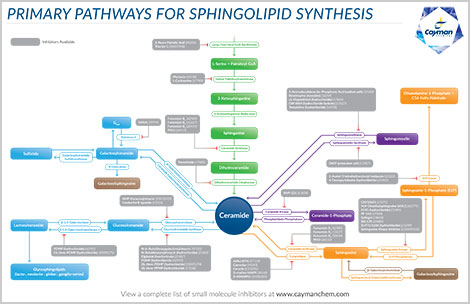
New Deuterium-Labeled Glycosphingolipids
Stable Isotope-Labeled Sphingolipids as Highly Specific Mass Spectrometry Standards
Novel Adamantane-Labeled Sphingolipids
Glycinated Lyso-Glycosphingolipids as New Mass Spectrometry Internal Standards
References
1. Han, X. and Jiang, X. A review of lipidomic technologies applicable to sphingolipidomics and their relevant applications. Eur. J. Lipid Sci. Technol. 111(1), 39-52 (2009).
2. Futerman, A.H. and Hannun, Y.A. The complex life of simple sphingolipids. EMBO Rep. 5(8), 777-782 (2004).
3. Maceyka, M., Milstien, S., and Spiegel, S. Sphingosine kinases, sphingosine-1-phosphate and sphingolipidomics. Prostaglandins Other Lipid Mediat. 77(1-4), 15-22 (2005).
4. Merrill, A.H., Jr., Sullards, M.C., Allegood, J.C., et al. Sphingolipidomics: High-throughput, structure-specific, and quantitative analysis of sphingolipids by liquid chromatography tandem mass spectrometry. Methods 36(2), 207-224 (2005).
5. Merrill, A.H., Jr. Sphingolipid and glycosphingolipid metabolic pathways in the era of sphingolipidomics. Chem. Rev. 111(10), 6387-6422 (2011).
6. Lipsky, N.G. and Pagano, R.E. Intracellular translocation of fluorescent sphingolipids in cultured fibroblasts: Endogenously synthesized sphingomyelin and glucocerebroside analogues pass through the Golgi apparatus en route to the plasma membrane. J. Cell Biol. 100(1), 27-34 (1985).
Contact Info
Cayman Chemical1180 East Ellsworth RoadAnn Arbor, Michigan 48108 USAToll Free: (800) 364-9897(USA and Canada only)Fax: (734) 971-3640


