
News & Announcements
Citrullination and Carbamylation in Inflammation and Autoimmunity: Key Biomarkers for Disease Detection
Article from 2021-04-27
Rheumatoid arthritis, psoriasis, systemic lupus erythematosus, multiple sclerosis, Alzheimer's disease, and cancer are just some of the disorders impacted by citrullination, a process in which protein arginine residues are converted to citrulline residues. Though citrulline is a non-standard amino acid that cannot be incorporated during protein synthesis, citrullination is a common post-translational occurrence associated with several important physiological processes. Unfortunately, dysregulated citrullination in the context of inflammation can result in excess citrulline residues, and citrullination of self-antigens is associated with autoimmune responses wherein antibodies to citrullinated protein antigens (ACPAs) are formed.1 A major source of ACPAs comes from insufficient clearance of cellular debris from apoptosis and NETosis, a unique mode of neutrophil cell death of which citrullination is an important part.2 During NETosis, fibrous networks of DNA fragments, citrullinated histones, and granule enzymes including neutrophil elastase, myeloperoxidase (MPO), and cathepsin G are extruded from neutrophils. These components form neutrophil extracellular traps (NETs) that are meant to catch and kill extracellular pathogens, yet improper management creates a means by which citrullinated and non-citrullinated antigens are externalized and potentially identified by immune cells as foreign substances.
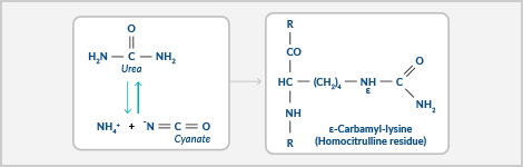
An additional protein modification very similar to citrullination is carbamylation. This nonenzymatic process involves the binding of cyanate (generated from urea dissociation or thiocyanate catabolism via MPO) to lysine residues resulting in the formation of homocitrulline. This reaction occurs at sites of inflammation, where thiocyanate is abundant in blood. For example, homocitrulline levels are especially elevated in smokers. The reaction is triggered by neutrophil-derived hydrogen peroxide and the MPO released in NETs. With just one additional methylene group, the homocitrulline residues formed by carbamylation are structurally similar to the citrulline residues formed by citrullination. Like ACPAs, autoantibodies to carbamylated proteins (anti-CarPs) are also generated. Anti-CarP autoantibodies and ACPAs show cross reactivity to some extent and frequently co-exist with each other.
Chemical reactions of citrullination and carbamylation
In a search for early identifiers of inflammatory or autoimmune diseases, attention has focused on detecting citrullinated and carbamylated proteins. For example, serum anti-CarP autoantibodies and ACPAs to several different antigens along with higher levels of NETosis are strongly associated with rheumatoid arthritis and other autoimmune disorders including systemic lupus erythematosus (SLE).3 Homocitrulline residues on proteins can indicate many of the same disease states as the presence of citrulline residues and in some cases reveal more severe disease progression.4 Autoantibody reactivity to citrullinated, carbamylated, and/or native proteins is linked to early stages of these diseases, making their utility as a biomarker intriguing for early diagnosis to improve therapeutic options. Ideally, if the profile of the disease is well known, multiple analytes, instead of just one biomarker, would allow for sufficient identification of the earliest disease onset, pinpoint the disease types and/or subtype, potentially predict the course of the disease or response to therapy based on known responses to treatment for that subtype, or even suggest relevant targets for prevention. Here we highlight several analytes of interest for autoimmune diseases.
Exposure to certain environmental or endogenous triggers leads to NET formation and increased PAD and MPO activity. Increased citrullination and carbamylation of peptides and proteins in the synovium creates new autoantigens that provoke an autoantibody response that becomes destructive to the surrounding tissues.
PAD Enzymes
The citrullination reaction is dependent on protein arginine deiminase (PAD) enzymes that deiminate arginine in a wide range of proteins in the cell membrane, cytoplasm, nucleus, and mitochondria. Five PAD isotypes (PAD1-4 and PAD6) exist in humans and have varied distribution, function, and pathology across the body. The irreversible actions of this enzyme result in a charge shift and a change in acidity that affects protein structure, protein-protein interactions, and hydrogen bond formation. This can lead to conformational changes that affect the binding and unfolding properties of the protein, which can ultimately alter its function and half-life. Protein denaturation can also occur. PADs are activated by relatively high calcium concentrations, and their function is important for normal cell processes such as skin keratinization and homeostasis, neuron insulation, gene regulation and expression, immune defense, and cell death. If PAD activities become uncontrolled, abnormal citrullination will lead to inflammation and disease. PAD enzymes along with citrullinated proteins can also leak from cells during massive cell death events that overwhelm the scavenger capacity of immune cells. This externalization of autoantigens can trigger autoimmune disorders.
Distribution, Targets, and Effects of PADs
| Isotype | Expression | Substrates | Physiology | Pathology |
| PAD1 | Epidermis, uterus, keratinocytes | Keratin K1, filaggrin | Epidermal tissue cornification | Psoriasis |
| PAD2 | Skeletal muscle, brain, secretory glands, inflammatory cells, mammary glands, uterus, cancer cell lines | Myelin basic protein, GFAP, vimentin, actin, histones H3/H4, fibrinogen, α-enolase | Myelin sheath stability, CNS plasticity, apoptosis, transcription regulation, innate immunity, fertility | Multiple sclerosis, Alzheimer's disease, prion disease, rheumatoid arthritis, SLE |
| PAD3 | Hair follicles, keratinocytes, neutrophils | Trichohyalin, filaggrin | Hair growth, epidermal function regulation, apoptosis | Unknown |
| PAD4 | Granulocytes, monocytes, macrophages, natural killer cells, stem cells, epithelial cells, tumors | Histones H2A/H3/ H4, myelin basic protein, inhibitor of growth 4 (tumor suppressor), p300/CBP (histone acetyltransferase), nucleophosmin/B23, nuclear lamin C, fibrinogen, thrombin, type II collagen, α-enolase | Chromatin decondensation, transcription regulation, tumor formation, innate immunity, apoptosis, NETosis | Rheumatoid arthritis, SLE, ulcerative colitis, multiple sclerosis, sepsis, thrombosis, cancer |
| PAD6 | Embryos, oocytes, ovary, testis | Protamine | Sperm chromatin decondensation, fertility, egg cytoplasmic sheath formation, early fetal growth, contraceptive drug target | Unknown |
In the case of rheumatoid arthritis, PAD2 and PAD4 are expressed in the synovium of the joint and catalyze the citrullination (often hypercitrullination) of arginine residues in proteins. The existence of these proteins has become a very important diagnostic feature for the disease.5 PAD2 and PAD4, which are overexpressed in immune cells including macrophages and neutrophils, are also relevant to many additional inflammatory diseases. The rheumatoid arthritis-associated citrullinome includes more than 150 citrullinated proteins identified through proteomic analysis, including fibrinogen, α-enolase, vimentin, filaggrin, keratin, β- and γ-actins, and histones that are then targeted for generation of ACPAs.6 These ACPAs begin to accumulate in rheumatoid arthritis patient serum 4-5 years before clinical onset of symptoms. Other autoantibodies that have been identified in these patients include antibodies against PAD enzymes themselves. Interestingly, a subpopulation of anti-PAD4 antibodies that show cross reactivity with PAD3 are strongly associated with joint erosions in rheumatoid arthritis patients.7 These autoantibodies that cross react between PAD3 and PAD4 increase the catalytic efficiency of PAD4 by reducing its requirement for calcium down into a more physiologic range.8 Because these autoantibodies activate an enzyme that then generates citrullinated autoantigens, a destructive feed-forward loop is created. Thus, the identification of PAD3 autoantibodies may provide a useful indication for the need for early aggressive treatment. One such therapy explored has been the use of PAD inhibitors in rheumatoid arthritis patients.
Histones
Histone modifications are central to the formation of NETs. Citrullination of histone tails results in chromatin decondensation and the lowering of the histone charge, which affects nucleosome stability and leaves DNA vulnerable to fragmentation. Neutrophils use this advantageously to fight infections by incorporating DNA fragments, citrullinated histones, and antimicrobial compounds from neutrophil granules, including the carbamylation catalyst MPO, into the NETs they release to initiate an immune response. But as discussed above, insufficient clearance of these components and the release of PAD and MPO into the extracellular space can contribute to inflammatory and autoimmune diseases. Autoantibodies to citrullinated histones have been detected in patients with rheumatoid arthritis and SLE. NETs also likely play a role in the homocitrulline-specific immune response in these diseases since histone carbamylation can occur at sites of inflammation.
Fibrinogen
Fibrinogen is another primary target for autoantibodies in the synovium of rheumatoid arthritis patients. As a terminal member of the clotting cascade, fibrinogen through the action of thrombin is converted to fibrin. It is highly upregulated during inflammation. Inflamed joints have excess deposition and local generation of fibrin that likely occurs due to imbalances between coagulation and fibrinolysis. Fibrinogen is citrullinated by PAD2 and PAD4 in synovial tissues. Carbamylated fibrinogen has also been identified.9 Thus, both citrullination and carbamylation of fibrinogen have a strong association with rheumatoid arthritis.
α-Enolase
α-Enolase is an additional, well-studied target of PAD2 and PAD4 in the rheumatoid arthritis synovium. The native form of α-enolase is a multifunctional protein, which catalyzes a step in glycolysis and also has roles as a cell surface receptor for plasminogen on pathogens, a structural lens protein, a tumor suppressor, and an autoantigen for several diseases. It is known to be upregulated by hypoxia and by pro-inflammatory stimuli, both of which are found in the rheumatoid arthritis synovial membrane microenvironment where citrullinated α-enolase has been detected. Antibodies targeting the citrullinated α-enolase are specific to rheumatoid arthritis.10 A bacterial enolase from P. gingivalis, a pathogen involved in periodontitis, has been linked to the formation of antibodies recognizing human citrullinated and native enolase and the rapid onset of arthritis.11 The sequence of the citrullinated peptide in human α-enolase is 92% homologous with its P. gingivalis counterpart. Interestingly, P. gingivalis also expresses its own unique PAD enzyme, which further implicates a pathogenic infection in the etiology of the disease.
Immunological Multiparameter Profiling
Because ACPA and anti-CarP autoantibody responses exhibit reactivity against multiple targets, multiplexed technologies are beneficial for the further understanding of their expression and disease association.12 In collaboration with Quansys Biosciences, Cayman created the Q-Plex™ Autoantibody Detection 10-Plex Panel as a qualitative ELISA that simultaneously measures human antibodies against PAD3, PAD4, fibrinogen, citrullinated fibrinogen, carbamylated fibrinogen, human core histones, citrullinated histones, carbamylated histones, α-enolase, and citrullinated α-enolase in plasma and serum samples. Concurrent analysis of these ten prominent analytes using a standardized assay platform creates a complex biomarker signature that could be mapped to disease types and subtypes. This approach is more robust and informative than single analyte detection, providing greater experimental control, more efficient use of labor efforts, and a more economical approach than running multiple single assays.
Q-Plex™ assay plates are built by adsorbing each distinct antibody or protein in a defined array to the bottom of each well. High-quality reagents provided in each kit help ensure the accuracy of the results. These assays require analysis using the Q-View™ Imager LS. This optimized imaging system is available from Cayman and includes a powerful, user-friendly image analysis software package that enables the user to capture, view, and analyze microplate images.
Products that support research on the role of citrullination and carbamylation in rheumatoid arthritis and other autoimmune diseases.
Generate an Experimental Autoimmune Disease Model
Immunizing mice with citrullinated fibrinogen induces the production of APCAs, citrulline-specific T cell activation, and a rheumatoid arthritis-like pathophysiology.13 The introduction of other citrullinated or carbamylated proteins is likely to have parallel effects. By promoting the production of ant ibodies that recognize citrullinated or carbamylated epitopes, this model may more closely approximate the pathophysiology of the human disease compared to other mouse models of arthritis, including collagen-induced arthritis.
Identify and Test
In the clinic, detection is important for early diagnosis and course of therapy. In animal models, detection is used to correlate degree of inflammation, PAD expression, citrullination, carbamylation, and autoantibody production with experimental variables. These tools will help detect citrullinated and carbamylated proteins and accurately analyze the antibody response induced in mice. Active recombinant PAD enzymes can be used for functional studies, PAD antibodies can be used for detection of the enzyme, and an assay is available to identify autoantibodies generated against PAD4 itself.
Develop Therapeutics and Screen for Efficacy
Reduction of citrullination is one therapeutic approach to ameliorate rheumatoid arthritis. However, caution must be taken with this approach since the physiological roles of PAD enzymes are quite important. Both selective and pan-PAD inhibitors are available that can be used as a companion to screening assays developed to identify novel inhibitors of PAD2, PAD3, and PAD4.
PAD Inhibitor Screening
PAD2 Inhibitor Screening Assay Kit (Ammonia)
PAD2 Inhibitor Screening Assay Kit (AMC)
PAD3 Inhibitor Screening Assay Kit (Ammonia)
PAD4 Inhibitor Screening Assay Kit (Ammonia)
PAD4 Inhibitor Screening Assay Kit (AMC)
Inhibitor | Target |
| BB-Cl-Amidine | PAD1-4 |
| Cl-Amidine (hydrochloride) | PAD1-4 |
| F-Amidine (trifluoroacetate salt) | PAD1 and PAD4 |
| BB-Cl-Yne | PAD1-4 (clickable) |
| BB-F-Yne | PAD1-4 (clickable) |
| CAY10723 | PAD2 |
| CAY10727 | PAD3 |
| CAY10740 (hydrochloride) | PAD4 |
| GSK106 (hydrochloride) | inactive control |
| GSK121 (trifluoroacetate salt) | PAD4 |
| GSK199 (hydrochloride) | PAD4 |
| GSK484 (hydrochloride) | PAD4 (reversible) |
| Photoswitchable PAD Inhibitor (technical grade) | PAD1-4 (photoactivated) |
| YW3-56 (hydrochloride) (technical grade) | PAD2 and PAD4 |
Investigate NETosis
PADs have been linked to inflammation through their role in catalyzing histone H3 hypercitrullination during NET formation. Cayman has developed a collection of tools to study this form of pathogen-induced cell death.
Isolate Neutrophils and Induce NET Formation Neutrophil (mouse) Isolation Kit Detect Neutrophil Elastase Neutrophil Elastase Activity Assay Kit | Visualize and Quantify NETs and Associated Enzyme Activity NETosis Imaging Assay Kit Detect MPO Neutrophil Myeloperoxidase Activity Assay Kit |
Related NET Inhibitors
Inhibitor | Target |
| 4-Aminobenzoic Acid hydrazide | MPO |
| AZD 9668 | Neutrophil elastase |
| BAY-678 | Neutrophil elastase |
| Diapocynin | NADPH-oxidase |
| GW 311616A | Neutrophil elastase |
| IM-93 | NETosis and ferroptosis |
| Neutrophil Elastase Inhibitor | Neutrophil elastase |
| Sivelestat (sodium salt hydrate) | Neutrophil elastase |
| SSR 69071 | Neutrophil elastase |
| Myeloperoxidase Inhibitor Screening Assay Kit | MPO |
Need Help Detecting NETs in Your Experiment?
Use our expertise to detect in vitro NET formation and screen for modulators
- High-content imaging
- Enzymatic detection
- Citrullination/carbamylation detection
- Inhibitor screening
You May Also Be Interested In
 |  |
|---|---|
| Quantifying NETs | Post-Translational Modification through Carbamylation |
 |  |
| Neutrophil Biology | Neutrophil Defensive NETworks |
 |  |
| Citrullination Research Tools | Screening Inhibitors of Citrullination |
References
1. Alghamdi, M., Alasmari, D., Assiri, A., et al., An overview of the intrinsic role of citrullination in autoimmune disorders. J. Immunol. Res. 2019, 7592851 (2019).
2. Demoruelle, M.K., Bowers, E., Lahey, L.J., et al. Antibody responses to citrullinated and non-citrullinated antigens in the sputum of subjects with and at-risk for rheumatoid arthritis. Arthritis Rheumatol. 70(4), 516-527 (2018).
3. Pruijn, G.J.M. Citrullination and carbamylation in the pathophysiology of rheumatoid arthritis. Front. Immunol.6, 192 (2015).
4. Shi, J., Knevel, R., Suwannalai, P., et al. Autoantibodies recognizing carbamylated proteins are present in sera of patients with rheumatoid arthritis and predict joint damage. Proc. Natl. Acad. Sci. U.S.A. 108(42), 17372-17377 (2011).
5. Aletaha, D., Neogi, T., Silman, A.J., et al. 2010 Rheumatoid arthritis classification criteria: An American College of Rheumatology/European League Against Rheumatism collaborative initiative. Arthritis Rheum. 62(9), 2569-2581 (2010).
6. Tilvawala, R., Nguyen, S.H., Maurais, A.J., et al. The rheumatoid arthritis-associated citrullinome. Cell Chem. Biol. 25(6), 691-704 (2018).
7. Seaman, A., Darrah, E., Infantino, M., et al. Anti-peptidyl-arginine deaminase 3 (PAD3) antibodies as a promising marker to measure joint damage in patients with rheumatoid arthritis. Autoimmun. Rev. 15(7), 776-780 (2016).
8. Darrah, E., Gile, J.T., Ols, M., et al. Erosive rheumatoid arthritis is associated with antibodies that activate PAD4 by increasing calcium sensitivity. Sci. Transl. Med .5(186), 186ra65 (2013).
9. Jones, J.D., Hamilton, B.J., and Rigby, W.F. Brief Report: Anti-carbamylated protein antibodies in rheumatoid arthritis patients are reactive with specific epitopes of the human fibrinogen β‐chain. Arthritis Rheumatol. 69(7), 1381-1386 (2017).
10. Kinloch, A., Tatzer, V., Wait, R., et al. Identification of citrullinated α-enolase as a candidate autoantigen in rheumatoid arthritis. Arthritis Res. Ther.7(6), R1421-R1429 (2005).
11. Lundberg, K., Kinloch, A., Fisher, B.A., et al. Antibodies to citrullinated α-enolase peptide 1 are specific for rheumatoid arthritis and cross-react with bacterial enolase. Arthritis Rheum. 58(10), 3009-3019 (2008).
12. Chandra, P.E., Sokolove, J., Hipp, B.G., et al. Novel multiplex technology for diagnostic characterization of rheumatoid arthritis. Arthritis Res. Ther. 13(3), R102 (2011).
13. Ho, P.P., Lee, L.Y., Zhao, X., et al. Autoimmunity against fibrinogen mediates inflammatory arthritis in mice. J. Immunol. 184(1), 379-390 (2010).
Contact Info
Cayman Chemical1180 East Ellsworth RoadAnn Arbor, Michigan 48108 USAToll Free: (800) 364-9897(USA and Canada only)Fax: (734) 971-3640


