
News & Announcements
Toll-like Receptors: Immune Signaling Sentinels
Article from 2021-03-18
Toll-like receptors (TLRs) are important innate immune proteins responsible for the identification of invading pathogens. They are pattern recognition receptors (PRRs) capable of recognizing specific pathogen-associated molecular patterns (PAMPs) conserved among microorganisms and also recognize damage-associated molecular patterns (DAMPs) from tissue injury or cell death resulting from inflammation, oxidative stress, necrosis, etc. Ultimately, TLRs act as sentinels, providing an immediate first line of detection of invading microbes leading to the activation of protective mechanisms of the innate immune system. They are also important for developing B cell- and T cell-mediated, pathogen-specific adaptive immune "memories." Cayman offers an extensive library of agonists, antagonists, and antibodies to study the function of these receptors. Tools to study TLR adaptor proteins and several downstream signaling effectors are also available. Read on to learn more about how they contribute to the TLR signaling pathway.
TLR Ligands
Ten human and 13 mouse TLRs specialize in identifying the molecular fingerprints of invading pathogens to activate an appropriate immune signaling pathway tailored to the nature of the infection. TLRs can distinguish among double-stranded and single-stranded RNA (dsRNA and ssRNA), unmethylated CpG DNA, bacterial lipopolysaccharide (LPS), lipoproteins, flagellin as well as synthetic analogs of various natural products (see Figure and TLR Ligand Table). TLR2 and TLR4 are the best characterized. TLR2 recognizes the bacterial cell wall components peptidoglycan (from Gram-positive bacteria), lipoproteins, and lipoteichoic acid (LTA) as well as zymosan and β-glucan from fungus. TLR4 recognizes LPS from Gram-negative bacteria. These PAMPs are accessible for TLR recognition once they encounter the cell surface. TLR4 is also activated by members of the S100 protein family, including S100A8/S100A9 (also called calprotectin) and S100A12 (also called calgranulin C), which act as DAMPs that promote inflammation. TLR1 and TLR6 form heterodimers with TLR2 for the discrimination of triacylated lipoproteins from diacylated lipoproteins, respectively. TLR5 is responsible for sensing flagella of motile bacterial species, and TLR11 recognizes pathogenic bacteria commonly associated with urinary tract infections such as uropathogenic E. coli, as well as a profilin-like protein from the parasite T. gondii. TLR10 is nonfunctional in mouse, but the HIV-1 glycoprotein gp41 has been identified as a human TLR10 ligand. TLRs 3, 7, 8, 9, and 13 recognize intracellular pathogen-derived nucleic acid motifs: dsRNA, ssRNA, ribosomal RNA (rRNA), or DNA delivered to intracellular compartments after the uptake of viruses and other pathogens or infected cells. TLR9 recognizes non-methylated CpG motifs of bacterial and viral DNA. Binding of ligand to TLR is thought to be unidirectional and irreversible, terminated only by endocytic removal from the cell membrane.
That's Weird! Curious about the name? These receptors were named for their similarity to the protein coded by the toll gene identified in Drosophila in the mid 80s by Christiane Nüsslein-Volhard and Eric Wieschaus, developmental biologists at the Max Planck Institute. The gene in question, when mutated, makes Drosophila embryos develop ventrally, when in normal development this should occur dorsally. As the story goes, the two researchers were so surprised when first viewing this unusual mutation from opposite sides of a double microscope that they both spontaneously looked up at one another and exclaimed "Das ist ja toll!", or "That's weird!". As it turns out, the toll gene is not only important for embryonic polarity during Drosophila development (a Nobel prize-winning discovery) but plays a key role in mammalian innate and adaptive immunity. |
TLR Specificity through Expression, Subcellular Localization, and Signaling
Specificity of TLR signaling comes through dimerization, adaptor combinations, downstream pathway bifurcations, and expression within specialized cell types. TLRs are expressed by various cell types including professional immune cells and Tregs (CD4+CD25+ regulatory T cells), as well as synovial fibroblast-like cells, epithelial cells, and all major glial cell types. The TLR family members are either expressed at the cell surface for extracellular ligand (lipid and protein PAMPs) recognition (these include TLRs 1, 2, 4-6, 11, and 12) or are localized in intracellular compartments such as endosomes, lysosomes, or endoplasmic reticulum for detecting bacterial and viral nucleic acid PAMPs (TLRs 3, 7, 8, 9, and 13). TLRs function through dimerization and oligomerization, associating with fellow TLRs or other co-receptors. Most TLRs appear to function as homodimers—though, as noted above, TLR2 forms heterodimers with TLR1 or TLR6—each dimer having different ligand specificity. TLRs may also depend on other co-receptors for full ligand sensitivity. For example, TLR4 recognition of LPS requires MD-2. CD14 and LPS-binding protein facilitate the presentation of LPS to MD-2.
TLR localization and signaling.
TLR signaling involves a sequence of stimulus-induced conformational changes in the receptor. Activation is a sequential process where ligand binding induces a conformational change in the C-terminal region of the extracellular domain, which stabilizes TLR-TLR interactions and, in turn, induces conformational changes in the TLR transmembrane domains. When a pathogen is identified by TLR receptor engagement, activated TLR recruits adaptor molecules within the cytoplasm of cells to propagate a signal. Four adapter molecules are known to be involved in signaling: MyD88 (myeloid differentiation primary response gene 88), TIRAP (Toll/IL-1 receptor (TIR) domain-containing adaptor protein), TRIF (TIR domain-containing adaptor-inducing IFN-β), and TRAM (TRIF-related adaptor molecule). These adaptors activate certain protein kinases (IRAK (interleukin-1 receptor-associated kinase) 1, IRAK4, TBK1 (TNF receptor-associated factor (TRAF) family member-associated NF-κB activator (TANK)-binding kinase), and IKK (IκB kinase)) that amplify the signal, leading to the induction or suppression of multiple genes that coordinate an inflammatory response to eliminate infectious agents.
All TLRs, apart from TLR3, recruit MyD88. Additionally, TLRs 1, 2, 4, and 6 recruit the additional adaptor CD14, which is required for LPS binding, and TIRAP, which links the conserved C-terminal intracellular TIR domain with MyD88. In the MyD88-dependent pathway, the MyD88 death domain recruits IRAK, which interacts with the adaptor protein TRAF6 and provides a link to the canonical NF-κB signaling pathway. Activation of the IKK complex consisting of IKKα, IKKβ, and IKKγ (also known as NEMO) targets the protein for ubiquitination and proteasome-mediated degradation, resulting in the release and nuclear translocation of NF-κB. The MyD88-dependent pathway also facilitates expression of MAPKs, such as p38 and JNK, and transcription factors, such as interferon regulatory factors (IRFs), influencing the production of a wide array of pro-inflammatory mediators, including reactive oxygen/nitrogen intermediates, cytokines, and chemokines.
TLR3 ligands initiate the TRIF-dependent pathway, whereas TLR4 can signal via either MyD88-dependent or TRIF-dependent pathways requiring the additional linker adaptor TRAM to associate with TRIF. In the TRIF-dependent pathway, TRIF interacts with TRAF3 to activate IRF3 and IRF7 and initiate IFN-α and IFN-β production, which are hallmarks of the host innate immune response to viral infection. Alternatively, TRIF can also bind to RIP1 (receptor-interacting protein 1) to initiate apoptosis or to activate NF-κB for late-phase induction of inflammatory gene expression. TLRs promote both cell death and cell survival signaling pathways. Cell survival pathways might be activated, for example, to prolong the lifespan of usually short-lived polymorphonuclear neutrophils to increase their effectiveness against pathogens, whereas cell death activation can be an effective means to kill microbes, preventing their spread throughout the rest of the organism. Because thousands of genes are activated by TLR signaling, the TLRs constitute one of the most pleiotropic yet tightly regulated gateways for gene modulation.
Cayman offers an extensive library of agonists, antagonists, and antibodies to study the function of these receptors. Use the tables below to navigate to research products associated with each TLR.
Related Inhibitors
Paquinimod—prevents S100A9 binding to TLR4
IRAK-1/4 Inhibitor—disrupts the activity of IRAK-1 and -4
TH1020—inhibits the TLR5-flagellin protein-protein interaction
Sophoradin—inhibits the TLR4 signal pathway at the level of NF-κB and MAPK
Related ELISA Kits
S100A8/S100A9 heterodimer (human) ELISA Kit
S100A12 homodimer (human) ELISA Kit
TLR Adaptor and Downstream Signaling Antibodies and Proteins
MyD88 (N-Term) Rabbit Monoclonal Antibody
IRAK-1 Polyclonal Antibody
IRAK-4 Polyclonal Antibody
TBK1 (human, recombinant)
TBK1 Monoclonal Antibody (Clone 4E6)
Cooperation between TLRs and Other PRRs
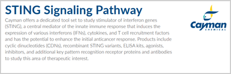
Non-TLR PRRs such as the cytosolic DNA sensor cyclic GMP-AMP (cGAMP) synthase (cGAS) and RNA sensors like the retinoic acid-inducible gene I (RIG-I)-like receptors (RLRs) offer an additional level of sophistication to the immune response that protects against viruses and bacteria. These sensors engage converging signaling cascades that lead to transcriptional induction of the genes encoding IFNs. Cytoplasmic DNA is sensed by cGAS and produces cGAMP. Stimulator of interferon genes (STING) detects cGAMP and relays the signal to TBK1 and IKKε. These in turn activate IRF3 and IRF7, which induce the expression of IFNs. In a similar way, cytosolic RNA is detected by RIG-I and other RLRs, which recruit the signaling adaptor protein mitochondrial antiviral-signaling protein (MAVS) anchored to mitochondria. Signaling through STING, MAVS relays the signal to TBK1 and IKKε, which activates IRF3 and IRF7 to induce the expression of IFNs. MAVS also stimulates NF-κB to induce the expression pro-inflammatory cytokines.
TLRs in Disease
Prolonged or excessive signaling through TLRs may lead to destructive inflammatory responses that ultimately result in exacerbating infection and undermining the very immune response that was intended to be protective. The presence of DAMPs, like viral ssRNA, can stimulate TLRs that activate resident immune cells to initiate and propagate inflammation and autoimmunity. Also, TLRs can recognize endogenous self-antigens and generate an aggressive autoimmune response in tissues. A crucial point in the generation of autoimmune disease is represented by defective apoptotic cell clearance that can lead to development of antinuclear antibodies. Inappropriate response of specific TLRs has been implicated in certain autoimmune diseases, immunodeficient diseases, inflammatory disorders, dementia, and cancer. For example, TLRs 2, 4, 7, and 9 participate in experimental allergic encephalomyelitis, a mouse model of multiple sclerosis, while TLR3 activation protects from this disease. TLRs 4, 7, and 9 have been connected to both human and mouse models of systemic lupus erythematosus and lupus-like syndromes. Furthermore, expression of TLRs 2, 4, 5, 7, and 9 is also increased in brain tissue of Alzheimer's disease patients and Alzheimer's transgenic mouse models. TLRs 3, 7, and 8 have important roles in the recognition of allergens and subsequently pathogenesis of allergic diseases as allergic rhinitis. Upregulation of some TLRs has also been shown in many tumor cells, tissues, and tumor cell lines.
Much is still to be determined toward understanding how TLR-mediated responses are tailored to produce either beneficial or detrimental effects, as well as how this signaling pathway can be harnessed for therapeutic benefit. Cayman's growing TLR product line contains several helpful research tools, including antibodies that recognize receptors and adaptor proteins.
You May Also Be Interested In
 STING Signaling Pathway Brochure | 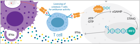 Helping Recruit Immune Cells to Cancer |
| | |
 Cyclic Dinucleotides: Ubiquitous Cellular Messengers |  Neutrophil Biology Wall Poster |
| |
Suggested Reading
Abreu, M.T. Toll-like receptor signalling in the intestinal epithelium: How bacterial recognition shapes intestinal function. Nat. Rev. Immunol. 10(2), 131-144 (2010).
El-Zayat, S.R., Sibaii, H., and Mannaa, F.A. Toll-like receptors activation, signaling, and targeting: An overview. Bull. Natl. Res. Cent. 43, 187 (2019).
Gambuzza, M., Licata, N., Palella, E., et al. Targeting Toll-like receptors: Emerging therapeutics for multiple sclerosis management. J. Neuroimmunol. 239(1-2), 1-12 (2011).
Golshiri-Isfahani, A., Amizadeh, M., and Arababadi, M.K., The roles of toll like receptor 3, 7 and 8 in allergic rhinitis pathogenesis. Allergol. Immunopathol. (Madr.)46(5), 503-507 (2018).
Hanke, M.L. and Kielian, T. Toll-like receptors in health and disease in the brain: Mechanisms and therapeutic potential. Clin. Sci. (Lond) 121(9), 367-387 (2011).
Hedayat, M., Netea, M.G., and Rezaei, N. Targeting of Toll-like receptors: A decade of progress in combating infectious diseases. Lancet Infect. Dis. 11(9), 702-712 (2011).
Henrick, B.M., Yao, X.-D., Zahoor, M.A., et al. TLR10 senses HIV-1 proteins and significantly enhances HIV-1 infection. Front. Immunol. 10, 482 (2019).
Kawai, T. and Akira, S. Toll-like receptors and their crosstalk with other innate receptors in infection and immunity. Immunity 34(5), 637-650 (2011).
Lepper, P.M., Triantafilou, M., O'Neill, L.A. Modulation of toll-like receptor signalling as a new therapeutic principle. Mediators Inflamm. 705612 (2010).
Marques, R. and Boneca, I.G. Expression and functional importance of innate immune receptors by intestinal epithelial cells. Cell. Mol. Life Sci. 68(22), 3661-3673 (2011).
Rehwinkel, J. and Gack, M.U. RIG-I-like receptors: Their regulation and roles in RNA sensing. Nat. Rev. Immunol. 20(9), 537-551 (2020).
Rozali, E.N., Hato, S.V., Robinson, B.W., et al. Programmed death ligand 2 in cancer-induced immune suppression. Clin. Dev. Immunol. 2012, 656340 (2012).
Siegmund-Schultze, N. Toll-like-Rezeptoren: Neue Zielstruktur für immunstimulierende Medikamente. Dtsch. Arztebl. 104(16), A-1072 / B-954 / C-908 (2007).
Contact Info
Cayman Chemical1180 East Ellsworth RoadAnn Arbor, Michigan 48108 USAToll Free: (800) 364-9897(USA and Canada only)Fax: (734) 971-3640


