
News & Announcements
How to Study Cellular Metabolism: Breaking Down Carbohydrate, Lipid, and Amino Acid Metabolism
Article from 2025-11-13
Cellular metabolism is a network of biochemical processes that occur in all cells to sustain life.1 These interconnected pathways fuel the bioenergetic and synthetic demands of all cellular functions and are coordinated to meet the cell's metabolic demand and availability of metabolic precursors.
Understanding alterations in cellular metabolism provides valuable insights into understanding disease mechanisms, drug effects on metabolism, cellular adaptations to environmental stressors, or preferential metabolic profiles in certain cell types.
Cellular Metabolism Research Tools Available from Cayman
Explore the assays and metabolic precursors, intermediates, and end products available from Cayman to study cellular metabolism.
Cellular Respiration
Cellular respiration is the major process that produces ATP, the primary source of energy for cellular processes.2 Cellular respiration is composed of three parts: glycolysis, the citric acid cycle, and oxidative phosphorylation.3
Glycolysis
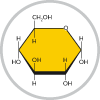
Glucose is a major substrate for cellular energy production. Glucose is obtained from dietary sources through the digestion of carbohydrates or from the hydrolysis of glycogen, a glucose storage form.4 Cellular uptake of glucose is mediated by glucose transporters (GLUTs), which transport glucose into the cytosol, where it undergoes a series of 10 enzymatic reactions to its final products.
Glycolysis produces two molecules of pyruvate for every one molecule of glucose.5 It requires two ATP molecules and two NAD+ molecules in the energy investment phase to produce a net gain of two ATP molecules and two NADH molecules during the energy payoff phase.
Overview of glycolysis. Glycolysis produces a net gain of two ATP molecules and two NADH molecules. For clarity, specific enzymes and metabolic intermediates are not shown.
Assays to Study Glycolysis
- Glycolysis Cell-Based Assay Kit
- Glucose Colorimetric Assay Kit
- Glucose Uptake Cell-Based Assay Kit
- Glycogen Assay Kit
- ATP Detection Assay Kit - Luminescence
- NAD/NADH Cell-Based Assay Kit
- Pyruvate Assay Kit
Anaerobic Glycolysis Research Tools
Notably, glycolysis is the only pathway that can generate ATP without oxygen, a process termed anaerobic glycolysis.4
During anaerobic glycolysis, pyruvate is converted to lactate in mammalian cells. This occurs in instances where oxygen cannot be delivered quickly enough to sustain high cellular metabolic demands, such as muscle cells during vigorous exercise. Under these conditions, NADH is used to reduce pyruvate to lactate, producing NAD+, which is used to fuel glycolysis.
In yeast, anaerobic glycolysis converts pyruvate to ethanol and carbon dioxide, reactions that are utilized in the production of alcoholic beverages.6
Overview of anaerobic glycolysis. Anaerobic glycolysis produces NAD+, which is essential to fuel glycolysis. Enzyme abbreviations: LDH, lactate dehydrogenase; PDC, pyruvate dehydrogenase complex; ADH, aldehyde dehydrogenase.
Assays to Study Anaerobic Glycolysis
- Pyruvate Assay Kit
- Ethanol Colorimetric Assay Kit
- L-Lactate Assay Kit
- D-Lactate Assay Kit
- NAD/NADH Cell-Based Assay Kit
The Citric Acid Cycle
The citric acid cycle, also known as the Krebs cycle or tricarboxylic acid (TCA) cycle, has a crucial role in cellular metabolism by capturing high-energy electrons from a variety of substrates.1 In the presence of oxygen, pyruvate enters the mitochondria, where it is oxidized to acetyl-coenzyme A (acetyl-CoA) by pyruvate dehydrogenase.4
Acetyl-CoA is condensed with oxaloacetate to form citrate, which initiates the citric acid cycle.1 Citrate undergoes a series of enzyme-catalyzed transformations to yield three NADH molecules, one FADH2 molecule, and one ATP (or GTP) molecule for every turn of the citric acid cycle. These reactions also regenerate oxaloacetate, refueling the citric acid cycle for further rounds.
Notably, the citric acid cycle does not directly produce substantial amounts of ATP.1 The citric acid cycle captures high-energy electrons from its substrates and transfers them to the electron carriers NADH and FADH2 to produce ATP during oxidative phosphorylation.
Overview of the citric acid cycle. The citric acid cycle produces a net gain of three NADH molecules, one FADH2 molecule, and one ATP (or GTP) molecule. For clarity, specific enzymes are not shown.
Assays to Study the Citric Acid Cycle
- MitoCheck® Citrate Synthase Activity Assay Kit
- Aconitase Assay Kit
- α-Ketoglutarate Detection Assay Kit
- ATP Detection Assay Kit - Luminescence
- NAD/NADH Cell-Based Assay Kit
Krebs Cycle Standard Mixture | |
A mixture of non-volatile acid Krebs cycle metabolic intermediates for GC-MS. Contains:
|
Oxidative Phosphorylation
Oxidative phosphorylation is the major process that produces cellular ATP, yielding 30 to 32 ATP molecules as compared to 2 ATP molecules from glycolysis alone.7
The electron transport chain is a series of protein complexes located in the inner mitochondrial membrane that transfers electrons from NADH and FADH2 to the electron shuttles coenzyme Q10, forming ubiquinol, and cytochrome c. As electrons move through the electron transport chain, their energy is used to pump protons out of the mitochondrial matrix and into the intermembrane space, creating a proton gradient. The proton pump creates an electrochemical gradient that drives protons back across the inner mitochondrial membrane through ATP synthase, the final protein complex in the electron transport chain, to form ATP.
Overview of the electron transport chain. The electron transport chain transfers electrons from NADH and FADH2 through protein complexes that establish a proton gradient. This proton gradient drives ATP synthesis, producing 30 to 32 ATP molecules.
Assays to Study Oxidative Phosphorylation
Electron Transport Chain
- MitoCheck® Mitochondrial (Tissue) Isolation Kit
- MitoCheck® Complex I Activity Assay Kit
- MitoCheck® Complex II Activity Assay Kit
- MitoCheck® Complex II/III Activity Assay Kit
- MitoCheck® Complex IV Activity Assay Kit
- MitoCheck® Complex V Activity Assay Kit
Electron Donors
Key Metabolic Product of Oxidative Phosphorylation
Proton Gradient
- JC-1 Mitochondrial Membrane Potential Assay Kit
- JC-1 Mitochondrial Membrane Potential Flow Cytometry Assay Kit
- TMRE Mitochondrial Membrane Potential Assay Kit
Oxygen Consumption Rate
Other Metabolic Pathways Intertwine with Cellular Respiration
Importantly, many of these reactions are reversible, and instead of performing catabolic functions, can also be used for anabolic activity, highlighting the versatility of the citric acid cycle under varying cellular conditions.1
Furthermore, depending on the metabolic requirements of the cell, many glycolytic and citric acid cycle substrates can be used in other pathways for the synthesis of other biomolecules, like NADPH, an electron carrier with important roles in redox homeostasis, or nucleotides, lipids, and proteins.4
The Pentose Phosphate Pathway
The pentose phosphate pathway both utilizes and produces glycolytic intermediates and is important for producing building blocks for nucleotide and amino acid synthesis and defense against oxidative stress.8 Thus, the pentose phosphate pathway links glycolysis, the citric acid cycle, and nucleotide synthesis.
The pentose phosphate pathway does not directly produce ATP; it produces NADPH and ribose-5-phosphate.9 The pentose phosphate pathway occurs in the cytosol in two phases: the oxidative phase and the non-oxidative phase. The oxidative phase uses glucose-6-phosphate, shunted from the glycolytic pathway, and converts it to 6-phosphoglucano-δ-lactonevia glucose-6-phosphate dehydrogenase (G6PD), the major rate-limiting step of the pentose phosphate pathway.10 This step produces NADPH, an electron carrier with important roles in redox homeostasis by regenerating reduced glutathione (GSH) from oxidized glutathione (GSSG) with additional roles in lipid synthesis. The oxidative phase ultimately produces ribulose-5-phosphate, a critical intermediate in nucleic acid synthesis.
During the non-oxidative phase, ribulose-5-phosphate is converted into ribose-5-phosphate, which can be used to produce 5-phospho-D-ribose 1-diphosphate (PRPP), a building block for purine and pyrimidine nucleotides.11 The non-oxidative phase also interconverts various sugars into glycolytic intermediates like glyceraldehyde-3-phosphate, which can be converted to pentose phosphates or re-enter glycolysis to form pyruvate.12
Overview of the pentose phosphate pathway. The pentose phosphate pathway produces NADPH, precursors for nucleotide synthesis, and glycolytic intermediates. Enzyme abbreviations: 6GPD, glucose-6-phosphate dehydrogenase; 6PGL, 6-phosphogluconolactonase; 6PGDH, 6-phosphogluconate dehydrogenase; GR, glutathione reductase; RPE, ribulose-5-phosphate-3-eipmerase; RPI, ribulose-5-phosphate isomerase; TKT, transketolase; TALDO, transaldolase.
Assays to Study the Pentose Phosphate Pathway
Assays for Redox Homeostasis
- NADPH:NADP Ratio Assay Kit
- Glutathione Reductase Assay Kit
- Glutathione Assay Kit
- Glutathione Peroxidase Assay Kit
Lipid Metabolism
Lipolysis
Lipolysis mobilizes the energy stored in triacylglycerols (TAGs) for cellular metabolism.13,14 TAGs are stored in lipid droplets and hydrolyzed into diacylglycerols (DAGs) and monoacylglycerols (MAGs) through sequential lipases to produce glycerol and free fatty acids. Glycerol can be used as a substrate for gluconeogenesis, and the free fatty acids can be converted to energy by either β-oxidation or the formation of ketone bodies.14,15
Overview of lipolysis. Lipolysis produces glycerol, which can be used as a substrate for gluconeogenesis, and free fatty acids, which are used for β-oxidation and ketogenesis. Enzyme abbreviations: ATGL, adipose triglyceride lipase; HSL, hormone-sensitive lipase; MAGL, monoacylglycerol lipase.
Assays to Study Lipolysis
- Lipid Droplets Fluorescence Assay Kit
- Adipolysis Assay Kit
- Triglyceride Colorimetric Assay Kit
- Lipase Activity Assay Kit
- Glycerol Colorimetric Assay Kit
- Glycerol Cell-Based Assay Kit
- Free Fatty Acid Fluorometric Assay Kit
β-Oxidation
Free fatty acids are transported in the circulation bound to albumin.16 Fatty acids bound to albumin are transported across the plasma membrane and then ligated to coenzyme A by acyl-CoA synthases. To enter mitochondria, the newly formed acyl-CoA must be conjugated to carnitine, an action performed by carnitine acyltransferases (CPTs). The acylcarnitine is then transported by carnitine acylcarnitine translocase (CACT) across the inner mitochondrial membrane.
View all acylcarnitines available from Cayman
The mitochondrial acylcarnitines are converted back to acyl-CoAs by CPTs. The resulting acyl-CoAs then undergo β-oxidation, a metabolic process that releases the two-carbon unit acetyl-CoA, NADH, and FADH2 during each cycle. Acetyl-CoA can enter the citric acid cycle, and NADH and FADH2 can be used for glycolysis and/or oxidative phosphorylation, all resulting in the production of ATP.17
Overview of β-oxidation. Free fatty acids are converted to acyl-CoAs, which must be converted to acylcarnitines to be transported to the inner mitochondrial membrane. The mitochondrial acylcarnitines are converted back to acyl-CoA, which undergoes β-oxidation to produce energy in the form of NADH, FADH2, and acetyl-CoA. For clarity, individual β-oxidation enzymes and metabolic intermediates are not shown.
Assays to Study β-Oxidation
Ketogenesis
Acetyl-CoA can also be used to produce ketones as an alternative energy source.18 Ketones are used by the body, especially in the brain, heart, and skeletal muscle, as an energy source when glucose is scarce.16 They are produced by the liver through ketogenesis, a process that occurs during periods of fasting, prolonged exercise, or low carbohydrate intake.18
During ketogenesis, ketones are formed by a series of reactions beginning with the condensation of two acetyl-CoA molecules by thiolase to form acetoacetyl-CoA, to which another acetyl-CoA molecule is ligated to form HMG-CoA.19 HMG-CoA is then cleaved by HMG-CoA lyase to produce acetoacetate, which is converted to β-hydroxybutyrate by D-βOHB dehydrogenase (BDH1).
Acetoacetate and β-hydroxybutyrate are transported to other tissues via the peripheral circulation, where they can be distributed to cells under metabolic demand. Once at their target cell, they are transported into the mitochondria where the reverse reactions occur, producing acetyl-CoA that can enter the citric acid cycle to produce ATP.
Overview of ketogenesis. Acetyl-CoA is used as a precursor to the formation of ketone bodies, which are used as an energy source during glucose scarcity. Enzyme abbreviations: THL, thiolase; HMGCS2, HMG-CoA synthetase 2; HMGCL, HMG-CoA lyase; BDH1, D-βOHB dehydrogenase.
Assays to Study Ketogenesis
- β-Hydroxybutyrate (Ketone Body) Colorimetric Assay Kit
- β-Hydroxybutyrate (Ketone Body) Fluorometric Assay Kit
Amino Acid Catabolism
One of the primary purposes of amino acids is protein synthesis. However, they can also be catabolized to produce a carbon skeleton that can be converted into products that are used for many metabolic processes, including citric acid cycle intermediates or as precursors for fatty acid synthesis and gluconeogenesis.20
View all amino acids available from Cayman
Glutaminolysis
Glutamine is the most abundant amino acid in the body.21 It is highly versatile, and glutamine can be used not only as a substrate for protein synthesis, but also for the synthesis of metabolic intermediates, nucleotides, and antioxidants.
Glutaminolysis is the metabolic pathway that catabolizes glutamine into several products that can be used for cellular metabolism and biological processes. Glutamine is converted by glutaminase to glutamate, a major excitatory neurotransmitter and precursor in glutathione (GSH) synthesis, which can be deaminated to form α-ketoglutarate, an important intermediate in the citric acid cycle that leads to the synthesis of ATP, and NADPH.22,23
Overview of glutaminolysis. Glutaminolysis produces glutamate, a neurotransmitter and precursor for GSH. Glutamate can be further metabolized to α-ketoglutarate, a citric acid cycle intermediate. Enzyme abbreviations: GLS, glutaminase; GDH, glutamate dehydrogenase.
Assays to Study Glutaminolysis
- Glutamine Fluorometric/Colorimetric Assay Kit
- Glutaminase Activity Assay Kit
- Glutamate Colorimetric/Fluorometric Assay Kit
- α-Ketoglutarate Detection Assay Kit
The Urea Cycle
Glutaminolysis (as well as catabolism of other amino acids) produces ammonia, a neurotoxic waste product that is converted in the liver to urea, which is excreted in the urine.3 Ammonia is produced in gaseous (NH3) and ionic (NH4+) forms that exist in equilibrium.4 Ammonia can either be converted back to glutamine when reacted with glutamate, an enzymatic reaction catalyzed by glutamine synthetase, or it can be detoxified in the liver via a series of biochemical reactions known as the urea cycle to produce urea, which is excreted in the urine.24
Overview of the urea cycle. Ammonia is detoxified to urea through a series of enzymatic biochemical reactions. Enzyme abbreviations: CPS, carbamoyl phosphate synthase; OTC, ornithine carbamoyltransferase; ASS, arginosuccinate synthase; ASL, argininosuccinate lyase; ARG, arginase.
Assays to Study the Urea Cycle
Markers of Cellular Metabolism
Cellular metabolism can also be studied or inferred by investigating indirect measures of heightened metabolic activity.
Proliferation
Cell division and growth is a metabolically intensive process. Accordingly, cellular proliferation can be used as a marker of cellular metabolism.
Assays
- XTT Cell Proliferation Assay Kit
- MTT Cell Proliferation Assay Kit
- WST-1 Cell Proliferation Assay Kit
- WST-8 Cell Proliferation Assay Kit
Immunochemical Markers
ROS
Reactive oxygen species (ROS) are a byproduct of cellular metabolism, making them a useful marker for increased cellular respiration.
- ROS Detection Cell-Based Assay Kit (DHE)
- ROS Detection Cell-Based Assay Kit (DCFDA)
- Mitochondrial ROS Detection Assay Kit
Protein Expression
Assessing expression levels of cellular energy sensors or metabolism-related proteins can also shed light on cellular metabolism.
Energy Sensors
Antibodies for Glycolysis Enzymes
- Glucose-6-phosphate Isomerase Monoclonal Antibody (Clone 5B6)
- α-Enolase Polyclonal Antibody
- GAPDH (C-Term) Rabbit Monoclonal Antibody (RM114)
Related Research Tools Available from Cayman
Protein Normalization Assays
Protein normalization assays adjust for variances in protein levels across samples, such as those observed in metabolically active cells or differences in cell numbers resulting from increased proliferation.
Mitochondrial Research Tools
Perform imaging studies or alter mitochondrial metabolism with these mitochondrial research tools.
- MitoLite™ Fluorescent Reagents
- Mitochondrial Uncouplers
- Small Molecule Activators
- Small Molecule Inhibitors
Cellular Metabolism Screening Library
A screening library of ~160 cellular metabolism modulators.
Contents include modulators of:
- Glycolysis
- Oxidative phosphorylation
- Pentose phosphate pathway
- Citric acid cycle
- Nucleotide and lipid metabolism
- Redox homeostasis
Cellular Metabolism Screening Services
Cayman's Cellular Metabolism Screening services provide a detailed view of cellular metabolic function.
Using platforms such as the Agilent Seahorse XF Pro Analyzer, our assays can be conducted using a variety of systems ranging from primary cells to isolated mitochondria and can be used to demonstrate changes in metabolic function or to de-risk potential toxins.
References
1. Arnold, P.K. and Finley, L.W.S. Regulation and function of the mammalian tricarboxylic acid cycle. J. Biol. Chem. 299(2), 102838 (2023).
2. Vercellino, I. and Sazanov, L.A. The assembly, regulation and function of the mitochondrial respiratory chain. Nat. Rev. Mol. Cell Biol. 23(2), 141-161 (2022).
3. Ahmad, M., Wolberg, A., and Kahwaji, C.I. Biochemistry, electron transport chain. In: StatPearls [Internet]. Treasure Island (FL): StatPearls Publishing (2025). Available from: https://www.ncbi.nlm.nih.gov/books/NBK526105/
4. Chandel, N. S. Glycolysis. Cold Spring Harb. Perspect. Biol. 13(5), a040535 (2021).
5. Chaudhry, R. and Varacallo, M.A. Biochemistry, glycolysis. In: StatPearls [Internet]. Treasure Island (FL): StatPearls Publishing (2025). Available from: https://www.ncbi.nlm.nih.gov/books/NBK482303/
6. Maicas, S. The role of yeasts in fermentation processes. Microorganisms 8(8), 1142 (2020).
7. Deshpande, O.A. and Mohiuddin, S.S. Biochemistry, Oxidative Phosphorylation. In: StatPearls [Internet]. Treasure Island (FL): StatPearls Publishing (2025). Available from: https://www.ncbi.nlm.nih.gov/books/NBK553192/
8. Stincone, A., Prigione, A., Cramer, T., et al. The return of metabolism: Biochemistry and physiology of the pentose phosphate pathway. Biol. Rev. Camb. Philos. Soc. 90(3), 927-963 (2015).
9. Ge, T., Yang, J., Zhou, S., et al. The role of the pentose phosphate pathway in diabetes and cancer. Front. Endocrinol. (Lausanne) 11, 365 (2020).
10. Tsouko, E., Khan, A.S., White, M.A., et al. Regulation of the pentose phosphate pathway by an androgen receptor-mTOR-mediated mechanism and its role in prostate cancer cell growth. Oncogenesis 3(5), e103 (2014).
11. Chandel, N.S. Nucleotide metabolism. Cold Spring Harb. Perspect. Biol. 13(7), a040592 (2021).
12. Patra, K.C. and Hay, N. The pentose phosphate pathway and cancer. Trends Biochem. Sci. 39(8), 347-354 (2014).
13. Cho, C.H., Patel, S.,and Rajbhandari, P. Adipose tissue lipid metabolism: Lipolysis. Curr. Opin. Genet. Dev. 83, 102114 (2023).
14. Sharma, A.K., Khandelwal, R., and Wolfrum, C. Futile lipid cycling: From biochemistry to physiology. Nat. Metab. 6(5), 808-824 (2024).
15. Edwards, M. and Mohiuddin, S.S. Biochemistry, lipolysis. In: StatPearls [Internet]. Treasure Island (FL): StatPearls Publishing (2025). Available from: https://www.ncbi.nlm.nih.gov/books/NBK560564/.
16. Longo, N., Frigeni, M., and Pasquali, M. Carnitine transport and fatty acid oxidation. Biochim. Biophys. Acta 1863(10), 2422-2435 (2016).
17. Talley, J.T. and Mohiuddin, S.S. Biochemistry, fatty acid oxidation. In: StatPearls [Internet]. Treasure Island (FL): StatPearls Publishing (2025). Available from: https://www.ncbi.nlm.nih.gov/books/NBK556002/.
18. Puchalska, P. and Crawford, P.A. Multi-dimensional roles of ketone bodies in fuel metabolism, signaling, and therapeutics. Cell Metab. 25(2), 262-284 (2017).
19. Fulghum, K., Salathe, S.F., Davis, X., et al. Ketone body metabolism and cardiometabolic implications for cognitive health. NPJ Metab. Health Dis. 2, 29 (2024).
20. Liu, X., Ren, B., Ren, J., et al. The significant role of amino acid metabolic reprogramming in cancer. Cell Commun. Signal. 22(1), 380 (2024).
21. Cruzat, V., Macedo Rogero, M., Noel Keane, K., et al. Glutamine: Metabolism and immune function, supplementation and clinical translation. Nutrients 10(11), 1564 (2018).
22. Plaitakis, A., Kalef-Ezra, E., Kotzamani, D., et al. The glutamate dehydrogenase pathway and its roles in cell and tissue biology in health and disease. Biology (Basel) 6(1), 11 (2017).
23. Barmore, W., Azad, F., and Stone, W.L. Physiology, urea cycle. In: StatPearls [Internet]. Treasure Island (FL): StatPearls Publishing (2025). Available from: https://www.ncbi.nlm.nih.gov/books/NBK513323/.
24. Jakhar, D., Sarin, S.K. and Kaur, S. Gut microbiota and dynamics of ammonia metabolism in liver disease. NPJ Gut Liver 1(11), 1-8 (2024).
Contact Info
Cayman Chemical1180 East Ellsworth RoadAnn Arbor, Michigan 48108 USAToll Free: (800) 364-9897(USA and Canada only)Fax: (734) 971-3640


