
News & Announcements
A Guide to Ferroptosis: Mechanisms & Research Tools
Article from 2025-04-08
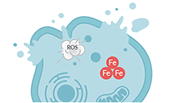
Ferroptosis is a form of cell death resulting from iron-dependent lipid peroxide accumulation. Iron triggers the peroxidation of fatty acids, resulting in the formation of highly reactive lipid peroxides. When these lipid reactive oxygen species (ROS) exceed the capacity of a cell's antioxidant system, the oxidative stress damages proteins, nucleic acids, and lipids, ultimately resulting in cell death.
Essential Ferroptosis Research Tools
Explore our quick product selections here or read on for in-depth product selections and information on why these factors are essential in studying ferroptosis.
Fundamental Biochemical Mechanisms of Ferroptosis
The interplay between iron availability, lipid peroxidation, and the production of ROS is a fundamental biochemical mechanism that drives ferroptosis.
| Target | Why It Matters | Detection Methods Available from Cayman |
| Iron Availability | Iron initiates lipid peroxidation and ROS formation via Fenton and Fenton-like reaction chemistries | Fluorescent Probes |
| Lipid Peroxidation | Iron-dependent lipid peroxidation damages cellular components | Assays Fluorescent Probes |
| ROS Levels | Excess iron promotes ROS formation through the Fenton reaction | Assays Fluorescent Probes |
Learn more about:
System xc-/GSH/GPX4 Axis
The system xc-/GSH/GPX4 axis is a critical pathway for ferroptosis that regulates intracellular glutathione (GSH) content to balance cellular redox.
| Target | Why It Matters | Detection Methods Available from Cayman |
| Cystine Uptake | A precursor in the formation of glutathione (GSH), an important mechanism for the detoxification of lipid peroxides | Assay |
| Glutamate Export | Intracellular glutamate is exchanged for extracellular cystine | Assays |
| Glutathione Redox | The GSH redox system detoxifies lipid peroxides, preventing ferroptosis | Assays |
| GPX4 Activity | GPX4 uses GSH to reduce lipid peroxides to less toxic products, preventing ferroptosis | Assays |
Learn more about the system xc-/GSH/GPX4 axis.
Ferroptosis Inducers
Several classes of small molecules can trigger ferroptosis by targeting lipid peroxidation underlying ferroptosis.
| Category | Mechanism | Effect |
| Class I FINs | System xc- inhibitors | Inhibits cystine uptake, resulting in GSH depletion and loss of GPX4 activity |
| Class II FINs | GPX4 inhibitors | Inhibits GPX4, inhibiting its antioxidant activity |
| Class III FINs | GPX4 depleters | Depletes GXP4, impairing its antioxidant activity |
| Class IV FINs | Iron oxidizers | Induces lipid peroxidation |
| FSP1 Inhibitors | FSP1 inhibitors | Inhibits FSP1reductase activity, impairing reduction of lipid peroxides |
Learn more about the factors that induce ferroptosis.
Ferroptosis Suppressors
Ferroptosis can be suppressed by inhibiting key initiators and propagators of the biochemical mechanisms underlying ferroptosis.
| Category | Mechanism | Effect |
| Antioxidants | Inhibiting the initiation and propagation of lipid peroxidation | Sequesters lipid peroxides, preventing cell damage |
| Iron Chelators | Binds and sequesters iron, preventing it from driving lipid peroxidation | Prevents the accumulation of excess iron |
Learn more about:
Factors that Induce Ferroptosis
Three key elements are needed to trigger ferroptosis: 1) polyunsaturated long-chain fatty acids (PUFAs) stored in phospholipid membranes, 2) a defective lipid peroxide repair system, and 3) redox-active iron.
Explore factors that contribute to ferroptosis with our lab wall poster.
Download or request a physical copy
1. Peroxidation of PUFA-PLs by LOs and PORs Drives Ferroptosis
Lipid Peroxidation & Oxidative Stress
PUFAs are highly susceptible to oxidative damage through both non-enzymatic and enzymatic processes (Figure 1). The bis-allylic hydrogens of PUFAs are prone to abstraction, which leads to the production of an alkyl radical (PL•) that readily reacts with O2 to produce peroxyl radicals (PLOO•) that react with other PUFAs to form phospholipid hydroperoxides (PLOOHs), creating a radical chain reaction of lipid peroxidation that results in ferroptosis.
Figure 1. Enzymatic and non-enzymatic mechanisms of ferroptosis.
| Top Selections: Tools for Lipid Peroxides & Oxidative Stress | |
|---|---|
| Lipid Peroxides: | |
| Assays View all lipid peroxidation assays | Probes |
ROS: | |
| Assays | Probes |
Radical-Trapping Antioxidants
Radical-trapping antioxidants (RTAs) inhibit ferroptosis by sequestering these radicals, preventing them from causing cellular damage.
| Top Selections: Radical-Trapping Antioxidants |
Mechanism
During ferroptosis, acyl-CoA synthetase long-chain family member 4 (ACSL4), lysophosphatidylcholine acyltransferase 3 (LPCAT3), lipoxygenases (LOs), and the cytochrome P450 oxidoreductase (POR) are the key drivers in the formation of oxidized PUFAs (Figure 2).
Figure 2. Enzymatic mechanisms of lipid peroxidation and ferroptosis.
Free intracellular arachidonic acid and adrenic acid are conjugated to coenzyme A (CoA) by ACSL4. LPCAT3 then catalyzes the esterification of these PUFAs into membrane phospholipids (PLs). LOs, particularly 15-LO, which normally uses free PUFAs as substrates, are then capable of oxidizing PUFA-PLH and generating PLOOHs, such as (e.g., PE-15-HpETE). If GPX4 inadequately reduces PLOOHs to PLOHs, the accumulation of PLOOHs will serve as a lethal signal to trigger ferroptosis.
Conjugated fatty acids (CFAs) also elicit ferroptosis-promoting effects, whereas the incorporation of saturated and monounsaturated fatty acids (SFAs and MUFAs) into cellular membranes counteract the effects of PUFAs and CFAs.
| Top Selections: Enzyme Inhibitors | ||
| ACSL4 Inhibitors | LPCAT3 Inhibitor | 15-LO Inhibitors |
2. Defective Lipid Peroxide Repair Mechanisms
The system xc-/GSH/GPX4 axis is a critical pathway for ferroptosis that regulates intracellular glutathione (GSH) content to balance cellular redox (Figure 3).
Figure 3.The system xc-/GSH/GPX4 axis controls GSH synthesis.
Cystine Depletion
System xc- transports intracellular glutamate to the extracellular space while transporting extracellular cystine into the cell.
| Top Selections: Tools to Study Cystine Depletion |
|---|
| Cystine Uptake Assay Kit A fluorometric, plate-based assay used to measure cystine uptake by culture cells View assay Class I FINs: System xc- Inhibitors These ferroptosis inducers interfere with GSH production by limiting cystine uptake. |
Regulation of System xc- Expression
Tumor suppressors p53 and BAP1 sensitize cells to ferroptosis by suppressing the transcription of a subunit of the system xc-, which reduces the import of cystine, and consequent synthesis of glutathione, a critical cellular antioxidant.
| Top Selections: Tools to Study Regulation of System xc- Expression |
| p53 Activators |
GSH Homeostasis
Cystine is transformed into cysteine for GSH synthesis. Under conditions of cysteine insufficiency, methionine can be converted to cysteine through the transsulfuration pathway to ultimately supplement the cysteine pool (Figure 4).
Figure 4. Methionine can be used as a precursor in the formation of cysteine, which is essential for GSH synthesis.
GSH is highly critical for protecting cells from damage caused by oxidative stress, and defective GSH synthesis increases the sensitivity of cells to ferroptosis due to disrupted oxidation-reduction balance.
| Top Selections: Tools to Study GSH | |
| Assays | GSH Depleters & Redox Modulators |
GPX4 Dysregulation
Glutathione peroxidase 4 (GPX4) is a lipid repair enzyme that utilizes GSH as a substrate to limit lipid peroxide accumulation (Figure 5). Thus, when the GSH pool is depleted, GPX4 activity is also reduced.
GPX4 is a selenoprotein that functions on biological membranes as a phospholipid hydroperoxidase. It catalyzes the reduction of lipid peroxides at the expense of GSH. When GPX4 activity is hindered, lipid peroxides accumulate and ultimately cause cell death.
| Top Selections: Tools to Study GPX | |
| Assays | Class II FINs These inducers suppress GPX4 activity, allowing lipid peroxides to accumulate. |
The Mevalonate Pathway & FSP1
Activation of squalene synthase (SQS) directs the MVA pathway towards cholesterol synthesis, suppressing the formation of several important non-sterol products like coenzyme Q10 (CoQ10), an important free-radical scavenging antioxidant (Figure 6).
Figure 6. The MVA and biopterin pathways play important roles in inhibiting ferroptosis.
Additionally, GTP cyclohydrolase 1 (GCH1) increases the de novo synthesis of CoQ10 by initiating the synthesis of dihydrobiopterin (BH2) and tetrahydrobiopterin (BH4). The actions of these proteins inhibit ferroptosis by acting as radical trapping antioxidants or contributing to the synthesis of reduced CoQ10.
The MVA pathway also plays a key role in GPX4 maturation, as isopentenyl pyrophosphate (IPP) contributes to the insertion of selenocysteine into the catalytic center of GPX4, which is important for its antioxidant activity. Loss of GPX4 function, either through loss of catalytic activity or by its degradation, promotes ferroptosis due to the loss of antioxidant activity.
| Top Selections: Tools to Study the Mevalonate Pathway | |
| Class III FINs This inducer negatively regulates GPX4 protein levels and activates squalene synthase (SQS). | Mevalonate Pathway Products |
FSP1
Ferroptosis suppressor protein 1 (FSP1) is an important regulator of ferroptosis that acts in a GSH-independent manner (Figure 7). FSP1 is an NADPH-dependent oxidoreductase that reduces CoQ10 to ubiquinol (CoQ10H2) and vitamin K to vitamin K hydroquinone (VKH2). The reduced forms of these molecules act as important reducing agents that are able to detoxify lipid peroxides.
| Top Selections: Tools to Study FSP1 | |
| Inhibitor Screening Assay Kit | FSP1 Substrates and Products |
| FSP1 Inhibitors | |
Regulation of FSP1 Expression
The MDM2/MDMX complex reduces expression of FSP1 by inhibiting PPARα activity.
| Top Selections: Tools to Modulate FSP1 Expression | |
| MDM2/MDMX inhibitors | PPARα Activators |
3. Iron Oxidation of PUFAs Initiates Ferroptosis
Regulation of Iron Levels & Metabolism
Excessive iron metabolism contributes to ferroptosis by producing oxidative stress. Circulating iron, in the form of ferric iron (Fe3+) bound to transferrin, is transported into cells through the membrane-bound transferrin receptor 1 (TfR1) (Figure 8). Within endosome compartments, Fe3+ is reduced to ferrous iron (Fe2+) by the metalloreductase STEAP3 and released into a labile iron pool in the cytoplasm via the divalent metal transporter 1 (DMT1) (Figure 4). Extra iron is stored in ferritin, an iron storage protein complex. Ferritinophagy and mitophagy also increase intracellular iron availability, promoting ferroptosis.
Any increase in iron uptake or reduced capacity for iron storage contributes to iron overload and the potential to generate highly reactive hydroxyl radicals through the Fenton reaction. These radicals can oxidize PUFAs in lipid membranes, creating lipid hydroperoxides.
| Top Selections: Tools to Study Iron Oxidation | |
| Class IV FINs These endoperoxides trigger ferroptosis by oxidizing Fe2+, which promotes lipid ROS. | Iron Chelators Iron chelation removes excess iron, preventing the formation of highly reactive hydroxyl radicals produced by the oxidation of Fe2+ in the Fenton reaction. |
| Iron Indicators | |
Metabolic Disturbances in Ferroptosis
E-Cadherin Signaling
Active proliferation is a marker of metabolically active cells. E-cadherin binding between cells mediates contact inhibition of proliferation when cells reach confluence (Figure 9). Contact inhibition of cell growth is essential for normal development, and uncontrolled cell growth is a characteristic of cancer. E-cadherin promotes contact inhibition through the Hippo signaling pathway. Binding of E-cadherin activates Hippo via NF2/Merlin, resulting in phosphorylation of YAP and TAZ, targeting it for proteasomal degradation and preventing their nuclear translation.
Figure 9. E-cadherin promotes contact inhibition of cell proliferation.
In the absence of Hippo signaling, YAP and TAZ are translocated to the nucleus, where they promote the activity of several genes involved in ferroptosis, including ACSL4 (e.g., lipogenesis) and the transferrin receptor (e.g., iron uptake).
| Top Selections: Proliferation Assays |
|---|
Energy Balance in Ferroptosis
mTORC1 and AMPK are master regulators of cellular energy status and metabolism that coordinate opposing functions. In energy-abundant conditions, mTORC1 is activated, whereas in low energy conditions, such as glucose scarcity, AMPK is activated. Both mTORC1 and AMPK can have pro- or anti-ferroptotic effects, depending on cellular context.
mTORC1
mTORC1 activation promotes the synthesis of proteins such as GPX4 and transsulfuration pathway enzymes (Figure 10). PI3K-Akt-mediated mTORC1 activation also encourages resistance to ferroptosis by promoting SREBP1-mediated increases in stearoyl-CoA desaturase 1 (SCD1), which catalyzes the conversion of SFAs to MUFAs.
Figure 10. mTOR activation promotes MUFA synthesis, inhibiting ferroptosis.
Accordingly, inhibition of mTORC1 is an intriguing strategy for sensitizing cancer cells to ferroptosis induction. However, mTORC1 inhibition has also been shown to inhibit ferroptosis by increasing levels of GSH.
| Top Selections: Tools to Study mTOR | |
|---|---|
| Inhibitors | Activators |
AMPK
AMPK signaling prevents ferroptosis by suppressing lipogenesis through inhibition of acetyl-CoA carboxylase (ACC), an enzyme responsible for the conversion of acetyl-CoA to malonyl-CoA, a key step in fatty acid synthesis (Figure 11).
Figure 11. AMPK signaling inhibits ferroptosis by downregulating the synthesis of new lipids.
This leads to decreased production of PUFAs, a major initiator of ferroptosis. On the other hand, AMPK activation downregulates MUFA production via SREBP, increasing susceptibility to ferroptosis.
| Top Selections: Tools to Study AMPK | |
|---|---|
| Inhibitors | Activators |
Mitochondrial Energy Production & Ferroptosis
Glutaminolysis
Glutaminolysis converts glutamine to glutamate, which fuels the TCA cycle. This increases the rate of oxidative metabolism in mitochondria, which causes the accumulation of ROS and promotes ferroptosis (Figure 12).
| Top Selections: Glutaminolysis Assay Kits |
|---|
Figure 12. Mitochondrial dynamics influence ferroptosis.
Mitochondrial ROS can hyperoxidize peroxiredoxin 3 (PRDX3), a thiol-based peroxidase with important roles in protection against oxidative stress. Hyperoxidized PRDX3 has been shown to block cystine uptake, limiting the synthesis of GSH and promoting ferroptosis.
| Top Selections: Tools to Study Mitochondrial ROS | |
|---|---|
| Assay | Probes |
| Detection Antibody | |
The Malate-Aspartate Shuttle
The malate-aspartate shuttle, which is important for providing energy for biosynthesis and maintaining redox balance, transfers reducing equivalents in the form of NADH from the cytosol into the mitochondria to facilitate oxidative phosphorylation (Figure X).
Disruption of glutamic-oxaloacetic transaminase 1 (GOT1), also known as aspartate aminotransferase (AST), within this shuttle has been implicated in creating metabolic vulnerabilities for cystine, glutathione, and lipid antioxidant function and has been exploited to trigger ferroptosis in cancer cells.
| Top Selections: Tools to Study the Malate-Aspartate Shuttle | |
|---|---|
| Assay | GOT1 Inhibitor |
Next Steps Towards Therapeutics
While important for controlling damage brought on by the oxidative stress response, ferroptosis has also been implicated in several pathological conditions in the brain, kidney, liver, and heart. This indicates a necessity for the development of therapeutics to inhibit the process. Indeed, much more work must be done to fully understand the underworking of ferroptosis. With expertise in bioactive lipid synthesis and protein, antibody, and assay development, Cayman is here to help in this endeavor by providing much-needed research tools.
Suggested Reading
Beatty, A., Singh, T., Tyurina, Y.Y., et al. Ferroptotic cell death triggered by conjugated linolenic acids is mediated by ACSL1. Nat. Commun. 12(1), 2244 (2021).
Bersuker, K., Hendricks, J.M., Li, Z., et al. The CoQ oxidoreductase FSP1 acts parallel to GPX4 to inhibit ferroptosis. Nature 575(7784), 688-692 (2019).
Chen, P.-H., Wu, J., Xu, Y., et al. Zinc transporter ZIP7 is a novel determinant of ferroptosis. Cell Death Dis. 12(2), 198 (2021).
Conlon, M., Poltorack, C.D., Forcina, G.C., et al. A compendium of kinetic modulatory profiles identifies ferroptosis regulators. Nat. Chem. Biol. 17, 665-674 (2021).
Doll, S., Freitas, F.P., Shah, R., et al. FSP1 is a glutathione-independent ferroptosis suppressor. Nature 575(7784), 693-698 (2019).
Gaschler, M.M., Andia, A.A., Liu, H., et al. FINO2 initiates ferroptosis through GPX4 inactivation and iron oxidation. Nat. Chem. Biol. 14(5), 507-515 (2018).
Jiang, X., Stockwell, B.R., and Conrad, M. Ferroptosis: Mechanisms, biology and role in disease. Nat. Rev. Mol. Cell Biol. 22(4), 266-282 (2021).
Kagan, V.E., Mao, G., Qu, F., et al. Oxidized arachidonic and adrenic PEs navigate cells to ferroptosis. Nat. Chem. Biol. 13(1), 81-90 (2017).
Kremer, D.M., Nelson, B.S., Lin, L., et al. GOT1 inhibition primes pancreatic cancer for ferroptosis through the autophagic release of labile iron. bioRxiv (2020).
* Lewerenz, J., Ates, G., Methner, A., et al. Oxytosis/ferroptosis—(re-) emerging roles for oxidative stress-dependent non-apoptotic cell death in diseases of the central nervous system. Front. Neurosci. 12, 214 (2018).
Ooko, E., Saeed, M.E.M., Kadioglu, O., et al. Artemisinin derivatives induce iron-dependent cell death (ferroptosis) in tumor cells. Phytomedicine 22(11), 1045-1054 (2015).
Shimada, K., Skouta, R., Kaplan, A., et al. Global survey of cell death mechanisms reveals metabolic regulation of ferroptosis. Nat. Chem. Biol. 12(7), 497-503 (2016).
Stockwell, B.R., Friedmann Angeli, J.P., Bayir, H., et al. Ferroptosis: A regulated cell death nexus linking metabolism, redox biology, and disease. Cell 171(2), 273-285 (2017).
Wenzel, S.E., Tyurina, Y.Y., Zhao, J., et al. PEBP1 wardens ferroptosis by enabling lipoxygenase generation of lipid death signals. Cell 171(3), 628-641 (2017).
Xie, Y., Hou, W., Song, X., et al. Ferroptosis: Process and function. Cell Death Differ. 23(3), 369-379 (2016).
Yang, W.S., Kim, K.J., Gaschler, M.M., et al. Peroxidation of polyunsaturated fatty acids by lipoxygenases drives ferroptosis. Proc. Natl. Acad. Sci. USA 113(34), E4966-E4975 (2016).
Yu, H., Guo, P., Xie, X., et al. Ferroptosis, a new form of cell death, and its relationships with tumourous diseases. J. Cell. Mol. Med. 21(4), 648-657 (2017).
Zhang, Y., Swanda, R.V., Nie, L., et al. mTORC1 couples cyst(e)ine availability with GPX4 protein synthesis and ferroptosis regulation. Nat. Commun. 12(1), 1589 (2021).
Zou, Y., Henry, W.S., Ricq, E.L., et al. Plasticity of ether lipids promotes ferroptosis susceptibility and evasion. Nature 585(7826), 603-608 (2020).
Zou, Y., Li, H., Graham, E.T., et al. Cytochrome P450 oxidoreductase contributes to phospholipid peroxidation in ferroptosis. Nat. Chem. Biol. 16(3), 302-309 (2020).
Contact Info
Cayman Chemical1180 East Ellsworth RoadAnn Arbor, Michigan 48108 USAToll Free: (800) 364-9897(USA and Canada only)Fax: (734) 971-3640


